Refinement of intraoral reverse temporalis transfer for facial palsy using a mandibular periosteum flap
Article information

Temporalis muscle transfer is an example of regional muscle transfer [1,2]. There are also ways to use a fascia lata graft to adjust the traction vector during muscle transfer [3]. This report describes temporal muscle transfer using the mandibular periosteum and partial temporalis detachment without a fascia lata graft to perform reconstruction in a patient with incomplete facial nerve palsy. First, the temporalis muscle was exposed through a temporal incision and the anterior third of the temporalis was detached from the temporal bone. Next, an intraoral incision was made to create an exposure from the mandibular coronoid process to the mandibular body. The periosteum was dissected from the mandibular body to the coronoid process connecting with the temporalis fascia. A saw was used to cut the coronoid process. The piece of the coronoid process connected to the temporalis was pulled to test whether the detached temporalis glided in the pulling direction. Maintaining the optimal shape of the nasolabial fold, the distal end of the periosteum was anchored to the muscle of the nasolabial fold through subcutaneous tunneling (Figs. 1, 2). The outcomes measured were preoperative and postoperative patient photographs (Fig. 3). The mouth corner showed improved symmetry in the resting state. Temporal area hollowness was not observed. The traditional temporalis muscle transfer technique uses a fascia lata graft to adjust the magnitude of the vector that exerts traction on the mouth corner. The technique introduced in this report enables adjustment of the magnitude of the vector applied to the mouth corner through partial temporalis detachment and by connecting the mandibular periosteum to the temporalis fascia. This technique produces satisfying cosmetic outcomes without the need for an additional donor site for fascia lata harvest.
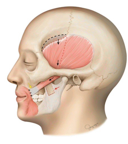
Schematic illustration of the surgical technique. The anterior third of the temporalis was gliding downward (black arrows) and the periosteum was dissected from the mandible (red arrow) and anchored to the muscle of the nasolabial fold (yellow arrow).
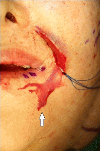
Intraoperative photograph. Distal end of the periosteum harvested from the mandible (white arrow); the proximal side was connected to the temporalis muscle.
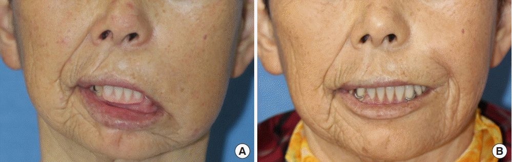
A 68-year-old woman with congenital incomplete left facial nerve palsy. (A) Preoperative resting state photograph. (B) Two months after the operation, in a resting state.
Notes
Conflict of interest
No potential conflict of interest relevant to this article was reported.
Ethical approval
The study was performed in accordance with the principles of the Declaration of Helsinki. Written informed consent was obtained.
Patient consent
The patient provided written informed consent for the publication and the use of her images.
Author contribution
Conceptualization: Sun H. Formal analysis: Han JW. Methodology: Han JW. Project administration: Han JW. Visualization: Han JW. Writing - original draft: Han JW. Writing - review & editing: Sun H.