Peptococcus Infection after Breast Augmentation Using Autologous Fat Injection
Article information
Autologous fat is widely used in reconstructive and aesthetic surgery [1]. Autologous fat insertion after a large amount of liposuction is possible, and autologous adipose injection has been widely used to restore dead spaces, correct scars, and improve facial or torso aesthetic contours.
However, there is ongoing discussion regarding the advisability of such surgery because the outcome of such a procedure is influenced by the method of fat collection, the amount of injected fat, the sites of fat collection and injection, and other unknown factors. The general complications associated with this surgery include edema, hematoma, infection due to fat necrosis or local reaction to the foreign substance, cyst formation, and irregular shape of the recipient site. Of these complications, the development of infection at the recipient site deserves the most attention. Abscess formation and in severe cases, sepsis, can threaten a patient's life [2]. We report a case of anaerobic infection that developed after breast augmentation surgery using autologous fat extracted from both the upper arms and abdomen.
A 39-year-old woman underwent autologous fat injection to correct defects in the upper and inner portion of both breasts using fat obtained through liposuction, 40 days before presentation at our hospital.
Her initial surgery was performed in an aesthetic plastic surgery clinic, and the amount of fat extracted and transplanted was not precisely known. Ten days before presentation at our hospital, the patient visited the clinic with a slight fever accompanied by edema, pain, and calor in the left breast. She received antibiotic treatment with ceftriaxone (CJ CheilJedang Corp., Icheon, Korea) and metronidazole (Kunhwa Corp., Gongju, Korea), but did not improve. At presentation, the swelling and calor in her left breast had worsened and both breasts had severe edema (Fig. 1).
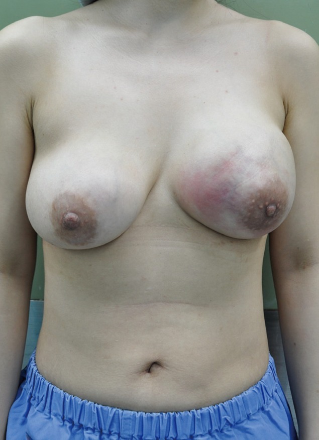
Preoperative photographic findings. Both breasts were swollen and the left breast showed cellulitic change with redness.
Blood tests showed an elevated C-reactive protein (CRP) level of 8.43 mg/dL, erythrocyte sedimentation rate (ESR) at 120 mm/hr, and white blood cell (WBC) count at 18,800 cells/µL. Emergency surgery was performed using a periareolar incision, and the abscess was drained. As the incisions were made, necrotic and melted fat that was seen as a clear, greasy, liquid discharge, which was followed by a yellowish discharge and finally a sticky, foamy discharge emerged. All were drained and removed together along with the necrotic tissue. The abscess in the left breast was drained of 120 mL of liquid, and 70 mL out of the right breast (Fig. 2). No foul odor was noted. The necrotic tissue and drained abscess contents were analyzed for the presence of gram-positive bacteria, gram-negative bacteria, anaerobic bacteria, and mycobacteria. An infection caused by gram-positive bacteria was assumed, and empiric vancomycin (2 g/day, CJ CheilJedang Corp.) was administered while waiting for the results of the culture and a consultation from an infectious disease specialist.
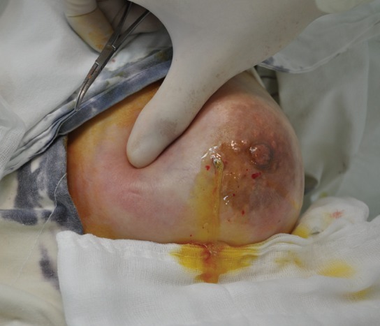
Intraoperative photographic findings. Drainage of a pus-like discharge with necrotic tissue.
The anaerobic bacterium, Peptococcus, was detected after 5 days of culture. Antibiotic treatment was continued until the 10th day of hospitalization. A follow-up blood test at that time showed normal levels of CRP (0.20 mg/dL), ESR (23 mm/hr), and WBC count (7,200 cells/µL). The patient's condition improved, and we discontinued antibiotic treatment based on the consultation from the infectious disease specialist.
The patient recovered fully. No complications such as recurrent infection or breast shape alteration were noted during follow-up. Symmetry and projection of the breasts were maintained. The patient was satisfied with her breast contour.
Autologous fat is considered an ideal material for expanding tissue because, unlike artificial implants, it is not associated with hypersensitivity or foreign body reactions. Autologous fat injection of the breasts is used for various purposes such as to correct imbalanced or protruding parts of the breasts. Autologous fat may also be used to reconstruct small defects that occur after breast cancer resection, shrinking of soft tissue following radiation treatment, or nipple reconstruction [3].
Complications such as fat tissue necrosis, scleroma, tiny calcifications, shape alteration or surface irregularity due to fat tissue absorption, and infection may occur. However, the most serious complication is the occurrence of infection. The inflammatory response of the recipient breast tissue to the transplanted fat can cause necrosis. Abscess formation is possible, and severe sepsis can be life threatening [2].
Bacteria that are commonly known to cause acute infections are Staphylococcus aureus (S. aureus), S. epidermidis, and S. pyogenes. All of these bacteria are sensitive to vancomycin; therefore, we empirically administered this antibiotic agent in this case. During emergency abscess drainage, a clear, oily discharge was first observed, followed by the yellow discharge of necrotic fat, and finally, a sticky, foamy discharge. The infected fat tissue was necrotic, and because it was surrounded by a belt of native fat, the abscess most likely formed from the inside. Interestingly, the bacterial culture obtained from the patient showed the presence of Peptococcus, which usually has a low pathogenicity and does not generally cause post-surgical infection. Peptostreptococcus is a genus of anaerobic, gram-positive, non-spore forming bacteria. The cells are small, spherical, and can occur in short chains, in pairs, or individually [4]. However, Peptostreptococcus is considered an important pathogen in the etiology of mixed anaerobic infections, and is usually absent or present in low numbers in the plaque of healthy individuals. We presumed that a synergistic infection was caused by a pathogenic aerobic gram-positive bacterium that is more frequently associated with post-surgical infections [5]; therefore, we continued the empiric treatment with vancomycin that targeted methicillin-resistant S. aureus after consulting with an infectious disease specialist. Because all of the signs and symptoms of infection improved within 10 days of incision and drainage along with vancomycin treatment, no additional antibiotic agent to specifically target anaerobic bacteria was administered.
The technique employed for collecting and injecting the fat is extremely important for a successful autologous fat transplantation. To reduce the possibility of complications, exposure of the fat to air before transplantation and mechanical damage should be minimized. An appropriate site should be chosen for the extraction of fat, according to the amount of adipose tissue needed, in order to minimize exposure of the collected tissue to the surrounding environment. Exact guidelines for successful fat transplantation surgery have not yet been established.
Notes
No potential conflict of interest relevant to this article was reported.