Hemangiopericytoma in the Nasolabial Fold
Article information
Hemangiopericytoma is rare in occurrence worldwide, including Korea. It was initially described by Stout and Murrary [1] in 1942. It is derived from a type of smooth muscle cell attached to pericytoma, capillaries also known as Zimmerman pericytes, and it is characterized by a "staghorn" shape on microscopic findings, in other words, a tumor with a diffuse pattern of branching, and dilated, thin-walled blood vessels surrounded by short spindle cells. Hemangiopericytoma has the very unusual characteristic of growing slowly, mainly in the lower extremities and pelvic retroperitoneum, but it can also be found in any part of the body such as the head and neck [1,2]. We introduce here a case of hemangiopericytoma found in the nasolabial fold because it rarely occurs in that area. Hemangiopericytomas in the head and neck are uncommon, but have been found in the neck, orbit, nasal cavity, parapharyngeal area, or tongue.
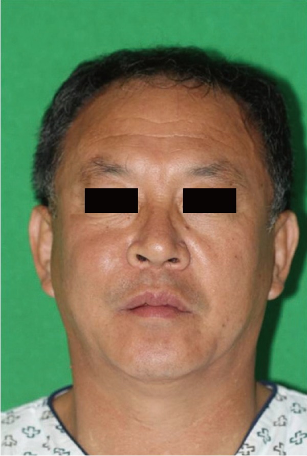
A 57-year-old male with no specific underlying disease was admitted to our hospital due to an increasing mass in the nasolabial fold as the chief complaint. The mass had been growing in size for 1 year. The mass was painless, non-fixed, movable, and round in shape. The mass grew in size until the nasolabial fold was more pronounced than the opposite side. The skin and buccal mucosa area around the mass were intact, and there was no drainage (Fig. 1). The computed tomography and magnetic resonance imagining findings showed a well-defined, solid mass with isoattenuation and heterogeneous enhancement in the premaxillary area (2.4×3.4×3.5 cm3) (Fig. 2).
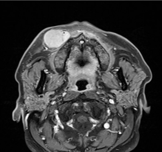
T1-weighted magnetic resonance imagining findings show a solid mass enhanced in the premaxillary area (2.4×3.4×3.5 cm3).
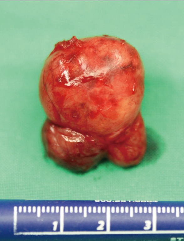
The operation was carried out under general anesthesia. The mass was large and considered to be a hemangioma or any other benign mass. However, the possibility of malignancy was not completely ruled out, and the incision was made through the nasolabial fold for minimal scarring. An approximately 5 cm incision was made along the right nasolabial fold, undermining the muscle layer. Complete excision of the mass was performed, and the mass was slightly irregular in shape and encapsulated, and was not attached to the surrounding tissues (Fig. 3).
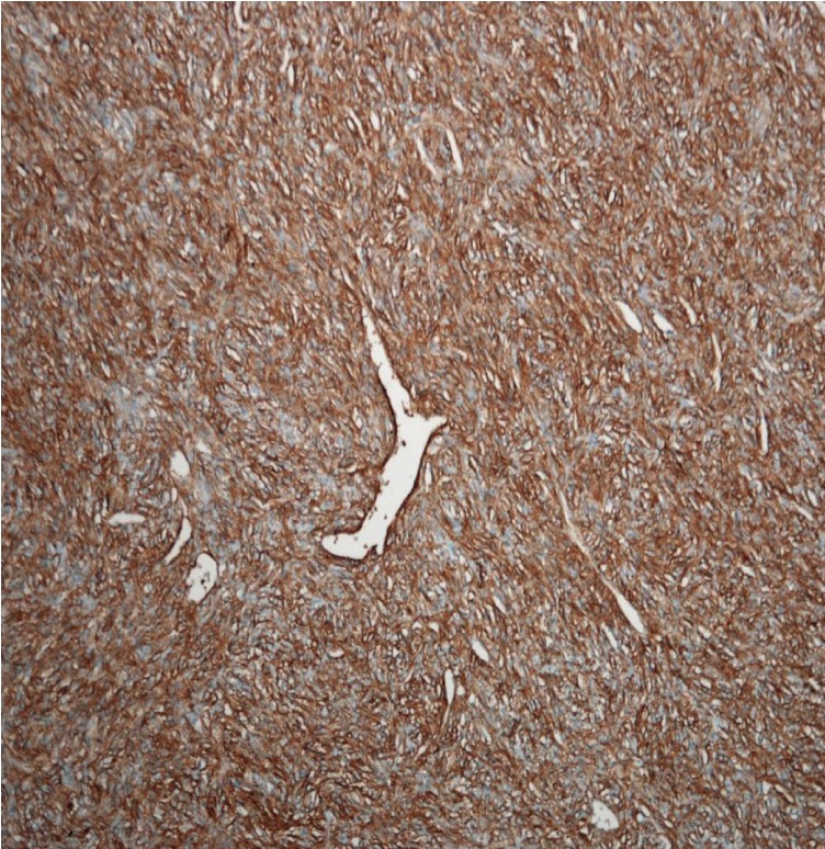
Microscopically, the tumor was composed of variably sized ectatic vessels showing a staghorn configuration and surrounding closely packed spindle or ovoid cells (Fig. 4). The tumor cells showed a small amount of pale cytoplasm and mild nuclear atypia, but had a low mitotic rate (less than 1 mitosis in 10 high power fields [HPF]). The tumor cells were diffusely positive in CD34 and CD99 immunostains (Fig. 5). There was no evidence of a tumor in the surgical margin. Postoperative chemotherapy and radiotherapy were not performed.
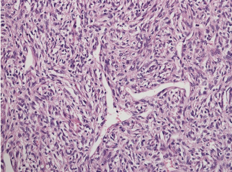
Tumor composed of ectatic, staghorn-shaped vessels and closely packed tumor cells, showing short spindle to round cells with a small amount of pale cytoplasm (H&E, ×200).
The patient did not mention any particular complaints for 3 months after surgery. There were no obvious facial changes or an awkward appearance when talking or showing emotions. The affected nasolabial fold had no significant difference from the opposite side. It seems that the dermal elasticity and subcutaneous adipose layer, which define the shape of the nasolabial fold, were not affected by the operation or the mass.
Hemangiopericytoma is characterized as a slow growing painless mass. When it occurs in the head and neck, patients are admitted to the hospital with symptoms caused by the mass physically affecting the surrounding tissues. The tumor is composed of spindle or round CD34 positive cells surrounding staghornshaped vessels. However, these features are overlapped with a solitary fibrous tumor; nevertheless, a solitary fibrous tumor has more prominent collagen, less prominent vessels, and less cellular pattern than a hemangiopericytoma. The clinical features of both tumors are also similar. Recently both types of tumors are considered to be the same entity with the two ends of one process. Hemangiopericytoma has a varied spectrum of biological behavior: benign to malignant. The prediction of the clinical behavior of hemangiopericytomas is not clear. Generally, a large size (>5 cm), increased mitotic rate (>4 mitosis/10 HPF) with the presence of atypical mitosis, high cellularity, pleomorphic tumor cells, and foci of hemorrhage and necrosis predict a highly malignant course [3,4].
The main treatment of hemangiopericytoma is complete surgical excision. Preoperative embolization can be helpful for decreasing the size of the mass. Radiotherapy, chemotherapy, or a combination of both can effectively improve the survival rates when the mass is inoperable or cannot be completely removed, or metastasis occurs.
Hemangiopericytoma has a varied spectrum of biological behavior: benign to malignant. Multiple articles have shown non-eventful life-long results after complete excision of hemangiopericytomas; however, it is better to consider a hemangiopericytoma to be malignant throughout a patient's lifespan because the tumor has the possibility of metastasis or recurrence. Therefore, long-term follow-up seems to be essential [4].
Notes
No potential conflict of interest relevant to this article was reported.
