Earrings Embedded within Earlobe Keloids
Article information
Keloids are a proliferative ailment of fibrous tissue secondary to dysregulation in various wound healing processes [1]. The diverse phenotypes and multitude of factors that trigger keloid formation have led us to propose "keloid disorder (KD)" as the identifying name for this condition and the term "keloid" to be reserved for referring to each individual skin lesion that patients have. Although benign, KD can cause aesthetic and functional problems, all of which pose a significant negative impact on the individual's quality of life. The earlobes are frequently involved sites for keloid formation following ear piercing, with an incidence of 2.5% [2]. Earlobe keloids are a cosmetic disfigurement that are challenging to treat with a relatively high recurrence rate. The increasing trend toward cosmetic piercing and multiple ear piercing suggests that treating ear keloids will become a more frequent part of plastic surgery practice. Diverse treatment modalities have been introduced with varying degrees of success. Various studies have estimated the onset of the disorder to be between 10 and 30 years of age [3].
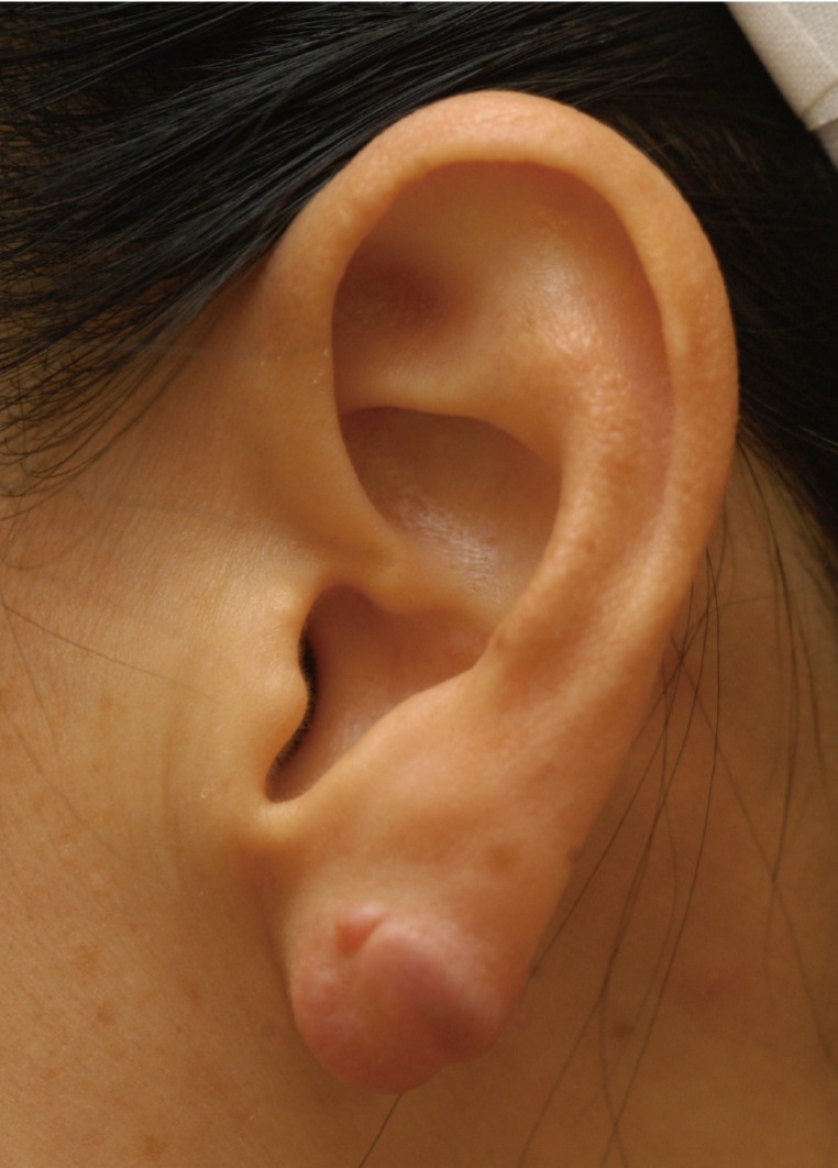
We have been faced with several earlobe keloids with earrings embedded within them. A representative case is presented in Figs. 1-3. Without exception, the cases in the series can be classified as having a sessile-type single nodular pattern based on our novel classification (Chang-Park classification) [4]. We completely excised the keloidal tissue, adopting full thickness wedge excision, which is considered to be the optimal treatment in this morphologic type. The wounds were closed with the appropriate approximation using nylon 5-0 continuous sutures. A compressive wound dressing using hydrocolloid materials and magnets was applied [5]. Following appropriate wound management, the patients were instructed to use the magnets for 12 hours per day for 6 months until the therapy was completed. The purpose of this report is to remind the reader that earrings may be embedded in earlobe keloids. Clinicians should keep this possibility in mind when faced with earlobe keloids.
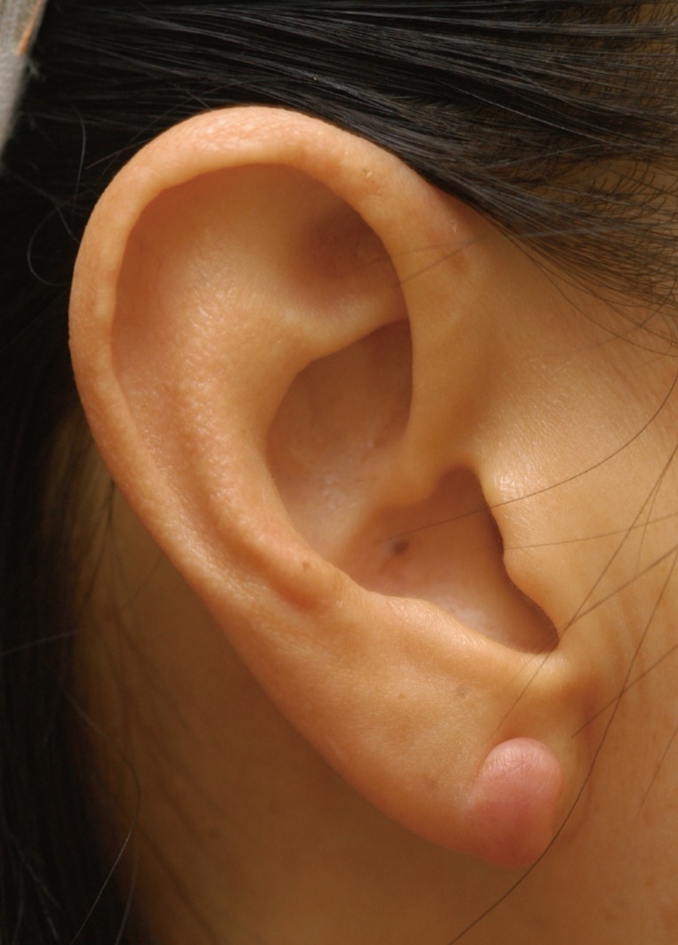
A right earlobe keloid; sessile-type single nodular pattern based on Chang-Park classification (type II).
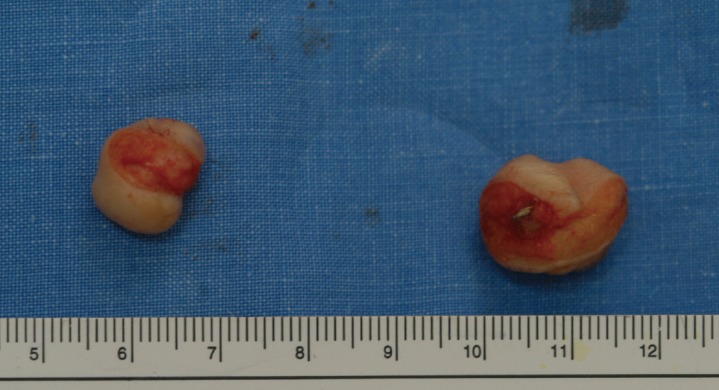
Earrings embedded within the earlobe keloids.
Notes
No potential conflict of interest relevant to this article was reported.