Correction of Severe "Pixie Ear" Deformity after Rhytidectomy with Modified Minimal Access Cranial Suspension Lift
Article information
The facelift remains the gold standard in facial rejuvenation, with a high satisfaction rate. However, excessive skin excision of the skin flap can produce unsatisfactory results and minor or even major complications. Amongst these complications, the pixie ear deformity can be recognized by its "stuck on" or "pulled" appearance, which is caused by the extrinsic pull of the medial cheek skin and jaw-line flaps at the earlobe attachment point, the otobasion inferius. Following rhytidectomy, the tension results in migration of the earlobe attachment point (otobasion inferius) from a posterior cephalad position to an anterior caudal position [1]. It is also possible that the pixie ear deformity can be accompanied by other tension-related complications such as a sweeping effect, scar migration, unnatural appearance of the tragus, and a "face-lift look."
In the medical literature, several techniques have been described for correction of this clinical condition. However, disadvantages such as extra scars on the neck or the earlobe and the controversial effectiveness of minimally invasive techniques in severe pixie ear deformities make the correction of this complication challenging to achieve. In this report, we present a case of severe pixie ear deformity after rhytidectomy, which was alleviated with a modification of the minimal access cranial suspension (MACS) lift.
A 53-year-old woman presented to our clinic totally dissatisfied with the postoperative result of a previous typical rhytidectomy performed elsewhere one year previously. Her main complaint was the unnatural appearance of the periauricular area. On clinical examination, severe pixie ear deformity and anterior migration of the preauricular scar were noted (Fig. 1). These facelift stigmata were the consequences of a rhytidectomy with overesection of the skin flaps, resulting in distortion of the position of the earlobe, which was attached to the angle of the mandible. With the patient under local anesthesia, we performed a simple MACS lift. The skin incision anterior to the earlobe was placed into the natural fold created after a slight anterior traction of the ear. A 5-cm undermining of the skin flaps was performed, and two loop-shaped sutures (polydioxanone, PDS 2-0) were applied and fixed to the deep temporal fascia. The first suture was anchored to the platysma, and the second to the superficial musculoaponeurotic system following the anterior border of skin undermining. The modification of the classic MACS lift was an additional PDS 2-0 purse-string suture, which was applied between the inferior border of the flap and the deep mastoid fascia in order to prevent the downward migration of the earlobe (Fig. 2). The undermined skin flap was elevated in a vertical vector and the redundant skin (about 1 cm in the upper part and 0.5 cm in front of the ear) was excised in order to achieve a tension-free skin closure. No suction drains were used. The postoperative period was uneventful, apart from a small blister that developed on the skin flap and mild erythema, which were resolved within the following days. The improvement of the earlobe position was found to have persisted at her follow-up visit after 18 months (Fig. 1).
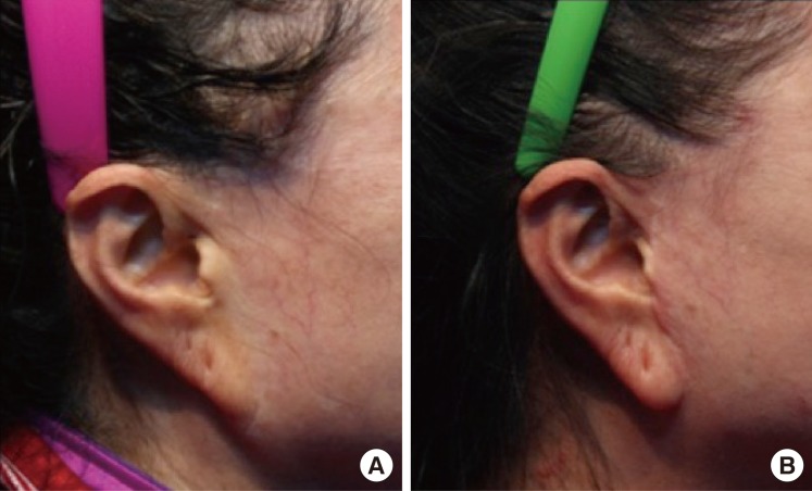
(A) Severe pixie ear deformity after a facelift. (B) Postoperative result at 18 months of follow-up.
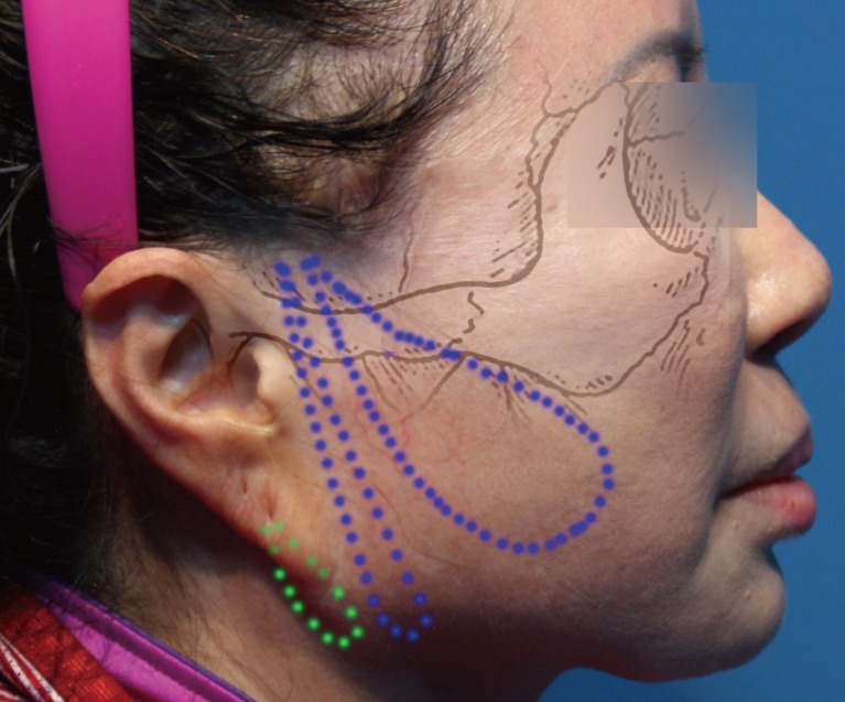
The first and second suture (blue dotted lines) were fixed to the deep temporal fascia 1 cm above the zygomatic bone and anchored to the platysma and the anterior border of the skin undermining, respectively. A third suture (green dotted line) was applied between the deep mastoid fascia and the inferior border of the flap. The deep mastoid fascia can be recognized as a stable structure after release of the anterior border of the earlobe.
In rhytidectomy, the skin closure under tension, especially in a pure horizontal vector, can lead to unsatisfactory results. Earlobe malposition, obvious scars, and an unnatural appearance of the tragus are complications whose improvement can be quite challenging due to the lack of available soft tissue. Amongst these complications, pixie ear deformity has been well described, and several techniques have been proposed to treat this condition.
Rich et al. [2] was the first to describe a minimally invasive technique to correct the pixie ear. After incision of the preexisting scars and the desired lobule borders, the deformed pointed lobule tip is excised and discarded, and the free margin of the lobule is approximated. Even though the technique is comprehensible and is able to correct severe deformities of the ear lobe, an extra scar is created vertically in the neck skin, making this method unattractive.
Mowlavi et al. [1] were the first to describe a method for correcting the pixie ear while avoiding the vertical scar in the neck skin. In their report, an algorithm is also presented for surgical correction of earlobe ptosis, pseudoptosis, and pixie ear deformity. Their technique was based on a wedge excision of the lobe, resulting in a horizontal scar on the lateral face of the lobe.
In 2001, Hoefflin [3] described a technique with almost invisible scars. After 3 stab incisions at the base of the lobule and undermining of the lobule, a 4-0 nylon suture is passed through the stab incisions, pulling the lobule superiorly. This methods looks ideal for minor pixie ear deformity; however, in their report neither long-term results nor cases with severe pixie ear deformity were presented.
Tepper and Zide [4] reported a novel method for correcting classic pixie ear or stuck-on ear after a facelift. Their technique is based on the phenomenon that the raw edges of the skin have a natural tendency to tube on themselves. In such cases, the earlobe is freed, the edge of the facial flap is readvanced, and the autotubing of the earlobe occurs within a few weeks. Even though the postoperative results are favorable, the usage of this method could be problematic in patients with abnormal wound healing and a tendency to develop hypertrophic scars or keloids.
The most recently presented technique belongs to Laurence et al. [5] from 2012. It relies on an asymmetric excision of an arrowhead-shaped wedge of tissue from the inferior-medial aspect of the helix, resulting in a hidden scar on the mastoid-facing portion of the newly formed lobe.
Our study involved a case of a woman with periauricular deformities following a facelift. Apart from the severe pixie ear deformity, an unnatural appearance of the tragus and anterior migration of the preauricular scar were the patient's complaints. During the consultation, it was found that the patient desired a minimally invasive procedure with no extra scars in the neck skin or the earlobe. With the patient under local anesthesia, we performed a simple MACS lift, applying an extra suture between the deep mastoid fascia and the skin flap. Elevating the skin flap to a pure vertical vector, we were able to reposition the earlobe to a higher location, recreating its normal curve, as well as to improve the tragus appearance and the preauricular scar. The support of the deeper tissues with sutures is critical to prevent recurrence.
The simplicity of the method, the absence of extra scars on the neck or the earlobe, and the satisfactory long-term results make the proposed technique very useful in improvement of post-rhytidectomy complications such as pixie ear deformity.
Notes
No potential conflict of interest relevant to this article was reported.