Thumb Carpometacarpal Dislocation and Open Dorsal Metacarpophalangeal Instability: A Variation of the Floating Thumb
Article information
With only five individual cases previously reported in the literature [1,2], the simultaneous dislocation of the carpometacarpal (CMC) and metacarpophalangeal (MCP) joints of the thumb without a fracture, also known as the floating thumb metacarpal, is an extremely rare injury.
We present a variation of this uncommon condition that was characterized by the presence of an open dorsal MCP instability and a concomitant CMC dislocation with no associated fractures. To the best of our knowledge, this is the first reported case of such a combination.
Our patient is a 59-year-old male who suffered direct trauma to the thumb after a fall on the sidewalk while drunk. He presented with pain and deformity at the base of the thumb and a 3-cm laceration on the volar aspect of the MCP joint.
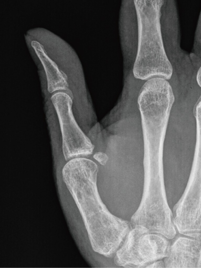
X-rays revealed a dorsal thumb CMC dislocation (Fig. 1). When examined under anesthesia, the MCP joint was also found to be dorsally unstable (Figs. 2, 3).
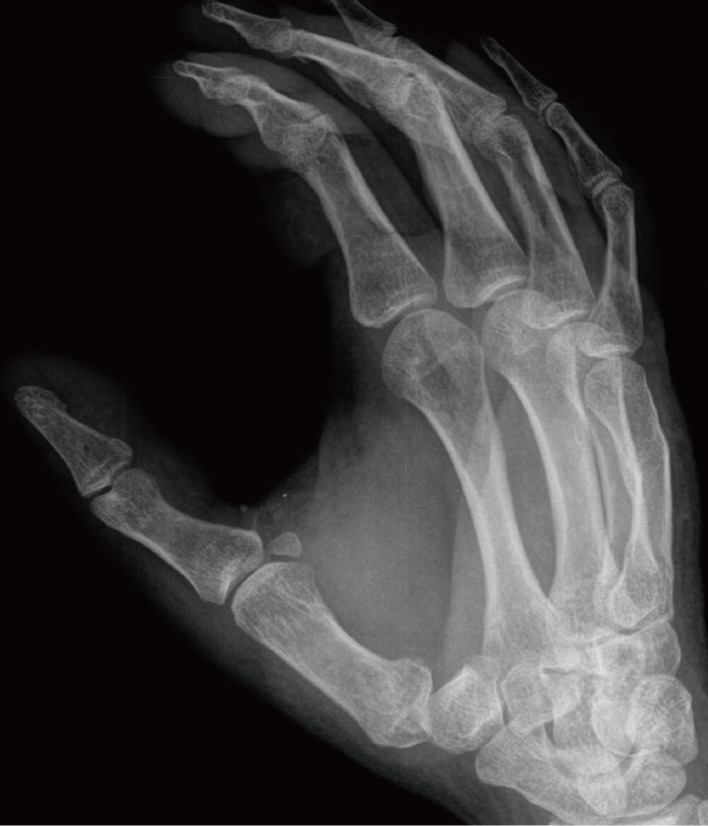
Left thumb carpometacarpal joint dislocation observed in X-rays.
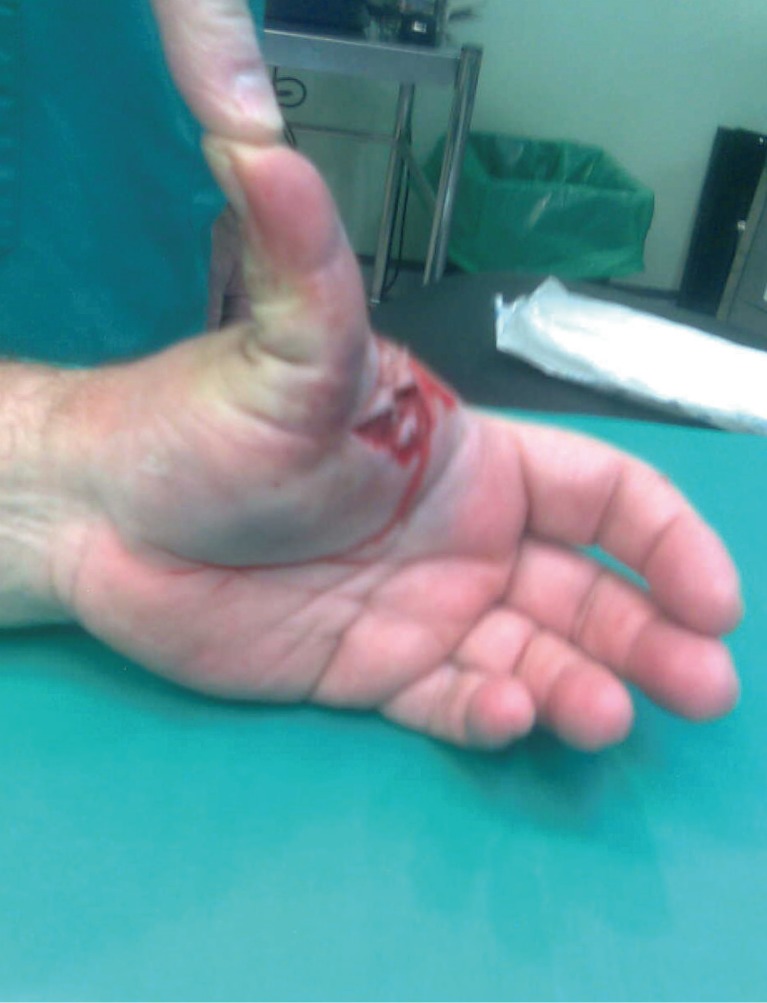
The metacarpophalangeal joint was easily dislocated when force was applied to the fingertip (lateral view).
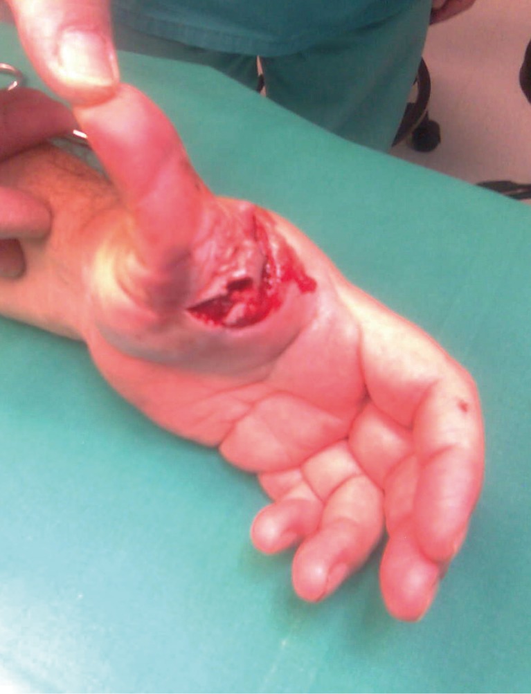
The metacarpophalangeal joint was easily dislocated when force was applied to the fingertip (frontal view).
The patient was taken to the operating room, where the CMC dislocation was reduced by closed manipulation and stabilized with two intermetacarpal Kirschner wires. The MCP injury was irrigated, and a significant tear of the volar plate was found to be the source of the instability; it was repaired with running stitches. An intra-articular Kirschner wire was used to further stabilize the joint (Fig. 4). A postoperative splint was then applied.
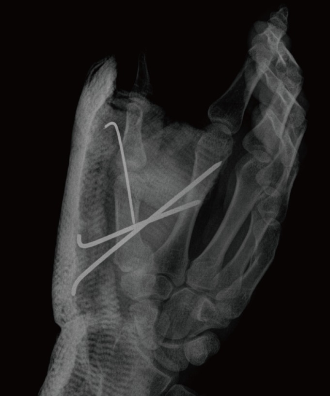
Immediate postoperative X-rays. Both joints are reduced and pinned with Kirschner wires.
The MCP wire was kept in place for 3 weeks, and the intermetacarpal wires were kept in place for 6 weeks, after which time, the splint was removed and active motion was encouraged.
After 34 months, the patient experienced some discomfort at the CMC joint. The range of motion at the interphalangeal joint was from 0° of extension to 45° of flexion; the MCP joint range of motion was from 0° of extension to 40° of flexion. Both the CMC and MCP joints were stable. Grip strength as measured with a Jamar dynamometer (Baseline Instruments, NY, USA) was 30 kg on the left and 35 kg on the right.
X-rays revealed osteoarthritic changes at the CMC joint (Fig. 5). The patient was satisfied with the outcome and wanted no further procedures.
After 34 months, incipient osteoarthritic changes were observed at the carpometacarpal joint.
Since the original description of this condition by Moore et al. [3] in 1978, five cases of floating thumb metacarpal have been reported in the literature [1,2].
A variation of this injury was previously reported by Bhargava and Jennings [4] in 2009. Their case involved a CMC dislocation and a simultaneous Stener lesion. The authors explained this as a result of a longitudinal force applied to the metacarpal, followed by radial stress to the MCP joint.
In our view, the present case is a result of a longitudinally directed force that dislocated the CMC joint and produced the open injury to the volar plate at the MCP level, resulting in a dorsal MCP instability.
Controversy remains as to the best treatment of CMC dislocations: closed reduction and casting, closed reduction and percutaneous pinning, and open reduction and ligament reconstruction have all been advocated, but no clear superiority of one technique has been clearly established [5]. Although no gross instability remained at the CMC joint in our case, some osteoarthritic changes developed over time.
In summary, we present a previously unreported variation of floating thumb, which was characterized by the combination of CMC dislocation and MCP instability; we thought that the injury combination was too unstable to be treated with reduction and casting alone and therefore, opted for stabilization with percutaneous pinning. In spite of the development of osteoarthritic changes at the CMC joint, the patient reported a minimal amount of pain and had a range of motion and grip strength that was comparable to that of the other thumb. We considered the clinical result to be a good one.
Clinicians should be aware that CMC dislocations can be associated with different types of MCP injuries, such as MCP dislocations, Stener-type lesions, or dorsal MCP instabilities. Diagnosis may be very clear or subtle. Treatment options include closed reduction and casting, closed reduction and percutaneous pinning, and open reduction and ligament reconstruction.
Notes
No potential conflict of interest relevant to this article was reported.