Incidental Total Necrosis of a Successful Flap Due to a Secondary Operation after One Year
Article information
Developments in microsurgical techniques and instruments have enabled the high success rate of microvascular tissue transfers. Initially, the survival of a free flap is dependent on its vascular pedicle, and the failure of flaps generally occurs within seven days of surgery. Several experimental studies have shown that pedicles can be ligated 5 to 10 days after the operation. Hence, at that time, the flap can be safely divided, and its survival can be ensured [1]. In addition, clinical studies have recommended that flaps can be safely divided between 7 and 21 days after surgery [2]. In this article, we report a case of the total loss of a flap after a secondary operation that was performed one year after successful reconstruction using a latissimus dorsi muscle flap.
A 68-year-old male presented with a chronic wound on his left leg. The patient had suffered an open fracture of his left tibia 15 years previously due to a fall. Radiologic findings revealed non-union of the tibia, and osteomyelitis was suspected. The orthopedic surgeon debrided the osteomyelitic bone, and we performed a reconstruction using a latissimus dorsi muscle free flap, which was 10 cm×8 cm in size, and a split thickness skin graft to cover the wound. The pedicle was located in the middle of the flap, and a posterior tibial vessel was used as the recipient vessel. The reconstruction was successful without complications such as partial necrosis, color change, or congestion (Fig. 1). Eighteen months later, the debrided bone with the bone defect seemed to be unstable, and the orthopedic surgeon planned an open reduction and internal fixation with a plate and free bone graft. A skin incision was made at the lateral margin of the flap, and the operation was conducted with a tourniquet applied. The location of the pedicle was marked using a hand Doppler before the operation, and no injury was found in the pedicle during dissection. There was no evidence that the main pedicle was bleeding after the tourniquet was removed. Immediately after the operation, the flap displayed a partial change of skin color (Fig. 2), and after three weeks, it became totally necrotic. We decided to discard the flap (Fig. 3) and reconstructed the defect with an anterolateral thigh flap, which was 12 cm×9 cm in size, by using the posterior tibial artery as the recipient vessel (Fig. 4). Three weeks later, the orthopedic surgeon removed the plate and the screws. A skin incision was made at the lateral margin of the flap. The method by which the perforator of the flap was marked was the same as before; the operation was successful, and no injury of the main pedicle during tourniquet application was observed. After the operation, the flap survived without complication, and the wound healed completely.
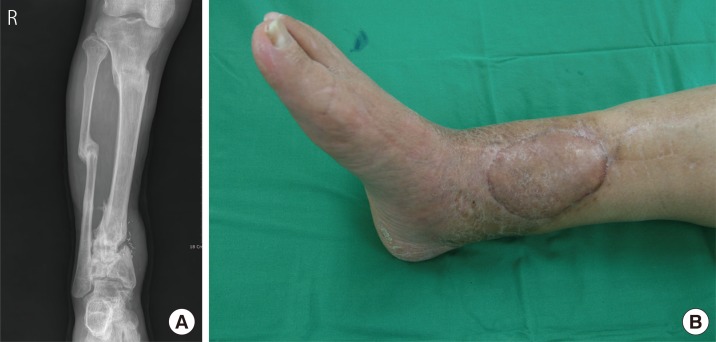
(A) A 68-year-old male presented with a chronic wound on his left leg. Radiologic findings revealed non-union of the tibia, and osteomyelitis was suspected. (B) The orthopedic surgeon debrided the osteomyelitic bone, and we performed a reconstruction using a latissimus dorsi muscle free flap and a split thickness skin graft to cover the wound.
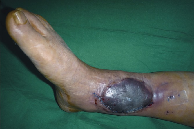
Immediately after the operation, the skin of the flap partially changed color.
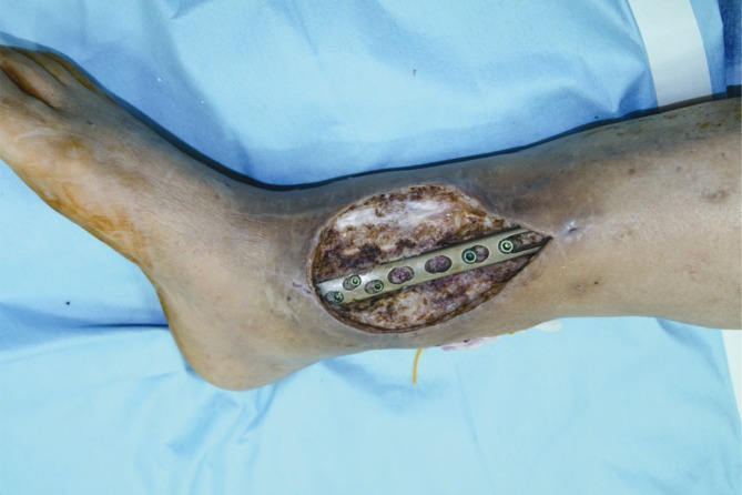
The flap was totally useless and hence, was discarded.
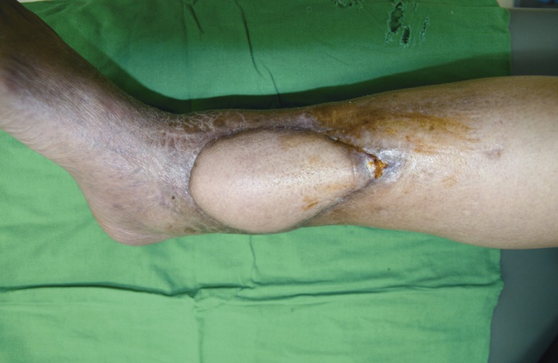
We reconstructed the defect with an anterolateral thigh flap using the posterior tibial artery as the recipient vessel.
After successful reconstruction by microvascular free tissue transfer, additional procedures are often performed. They include flap division to deal with multiple defects, liposuction for volume reduction, and debulking procedures for contouring. In our case, an incision was made at the flap margin, and the flap was lifted since the orthopedic surgeon required a secondary operation. Such additional procedures are made possible by neovascularization across the free flap from the surrounding tissue and from the wound bed; therefore, the flap is not totally dependent on the original pedicle for vascularity.
Experimental studies have demonstrated that neovascularization develops within 7 to 10 days [1]. On the other hand, there are a few case reports of flap failure as late as 16 months postoperatively [3], which suggests that there can be a significant blood supply from the anastomosed vessel even 1 or 2 years after surgery [4].
From these studies, it seems likely that most free flaps are neovascularized around seven days after the operation. However, several factors can lead to unpredicted late flap failure. Flaps may depend longer on the main vascular pedicle in patients with compromised recipient beds. Moreover, when the wound is chronic, the patient has peripheral vascular disease that affects the marginal blood supply, there is a subclinical infection, or the wound has received radiation treatment, then neovascularization of the flap is inhibited, development of a collateral circulation from the surrounding tissues or from the wound bed is hindered, and the flap remains dependent on the main pedicle [5]. In our case, the patient had suffered from a chronic wound for 15 years, which led to fibrous scarring that repeatedly healed and worsened, disrupting the peripheral vascularity.
It is well known that smoking affects the survival of free flaps and replantations by compromising vascularity. Our patient was a heavy smoker and continued smoking even after the operation. This may have affected neovascularization or capillary perfusion, even though the flap survived with the blood supply from the main pedicle.
In addition, although the skin incision was made at the lateral border of the flap, the flap itself was lifted completely, leaving only the opposite margin intact. Moreover, the flap might have been pulled and stretched during the operation to obtain a better view of the tibia. The previously formed neovascularization from the skin incision site and the wound bed may have been cut off, and the internal fixation with a plate and bone graft may have worsened the state of the wound bed. A plate and bone graft beneath a flap pushes on the flap, increasing the tension at its margin, and this may have affected the vascularity of the flap.
Although most free flaps are believed to be neovascularized around seven days after surgery, in one study, no new vessel formation was observed in the margins of the flap after the occlusion of the original anastomosis, and there was significant blood flow into the flap through the anastomotic vessels even after 1 to 2 years [4]. Therefore, additional procedures after free flap surgery should be approached with care, and either neovascularization across the flap should be confirmed or the main vascular pedicle should be left intact.
We report a case of the total loss of a flap after a secondary operation was performed 18 months after successful reconstruction using a latissimus dorsi muscle flap. There is controversy concerning flap neovascularization, and many factors can compromise flap vascularity including the flap itself, patient comorbidities, and the wound condition. Given these risks, additional procedures affecting the flap should be performed with considerable care, with attention paid to flap vascularity.
Notes
No potential conflict of interest relevant to this article was reported.