Macrodystrophia lipomatosa of bilateral hands and the left upper limb with syndactyly
Article information
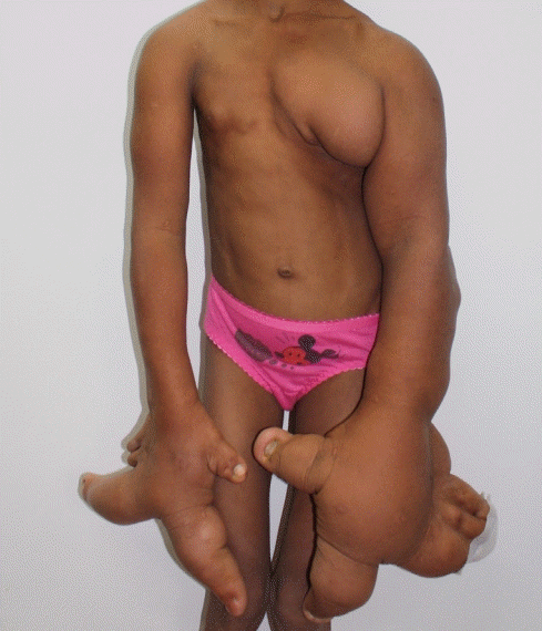
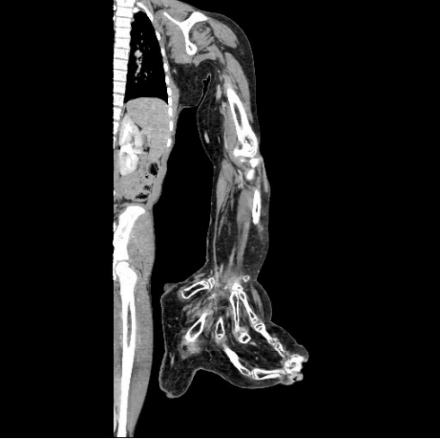
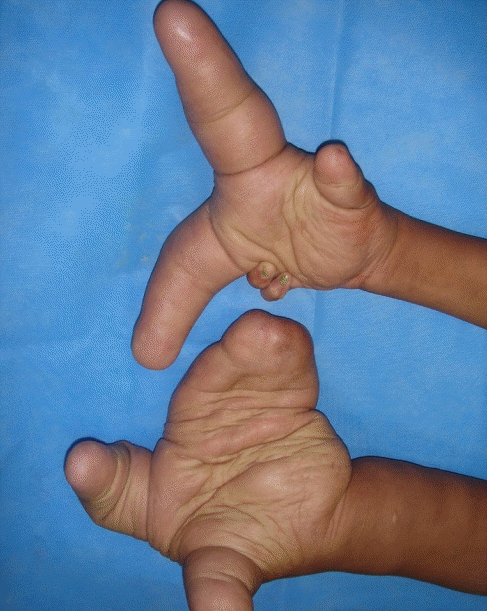
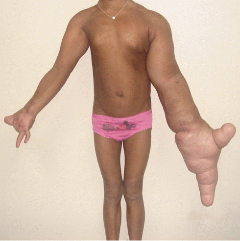
A 15-year-old girl from Myanmar presented to our department, and large growing malformations of the left upper limb and the dorsal (Fig. 1) and volar (Fig. 2) aspects of both hands were noted. These deformities had been present since birth, and they had progressively become more pronounced. The metacarpal and phalangeal bones of the right thumb, index, middle fingers and all the left fingers appeared hypertrophic on an X-ray examination. Computed tomography (CT) (Fig. 3) and magnetic resonance imaging (MRI) showed diffusely inflated fat tissue spreading through the lesions without encapsulation. Macrodystrophia lipomatosa (MDL) was diagnosed based on the patient’s clinical presentation and CT and MRI findings. The bilateral distribution of MDL, including the entire upper extremity, was extremely rare [1,2]. She underwent three operations. The first operation consisted of ray amputation of the right index and middle finger (790 g) and opponensplasty using the third flexor digitorum longus tendon. The second operation consisted of resection of the left axillary mass (370 g) and liposuction of the left chest wall and upper arm (250 mL). The third operation consisted of resection of lipomatous tissue in the left forearm with ray amputation of the left syndactylized middle, ring, and little fingers (900 g). Histopathology showed mature adipose tissue scattered within fine, mesh-like fibrous tissue. The wounds healed uneventfully. After surgery, she showed improved grasp in the right hand and reduced deformities of both hands and the left axilla (Fig. 4). The patient was no longer able to perform fine motor actions.

A 15-year-old girl showed a massive mass in the both hands and left upper limb. Her body posture was tilted by the asymmetrical weight of both upper limbs.

Computed tomography finding in the left upper limb shows enlarged adipose tissue spreading through the lesion without encapsulation.
Notes
No potential conflict of interest relevant to this article was reported.
Ethical approval
The study was approved by the Institutional Review Board of Bestian Seoul Hospital (IRB No. 2019-02-001) and performed in accordance with the principles of the Declaration of Helsinki. Written informed consent was obtained.
Patient consent
The patient provided written informed consent for the publication and the use of her images.


