Subtotal extended thigh flap
Article information

In 2010, Chang et al. [1] reported closing a wound after hemicorporectomy using a bilateral subtotal thigh flap. We describe a subtotal extended thigh flap for a 42-year-old male quadriplegic patient who presented with a nonmetastatic Marjolin ulcer (Fig. 1). Magnetic resonance imaging revealed that soft tissues, the coccyx, and the sacrum bone up to the S2 vertebra were involved.
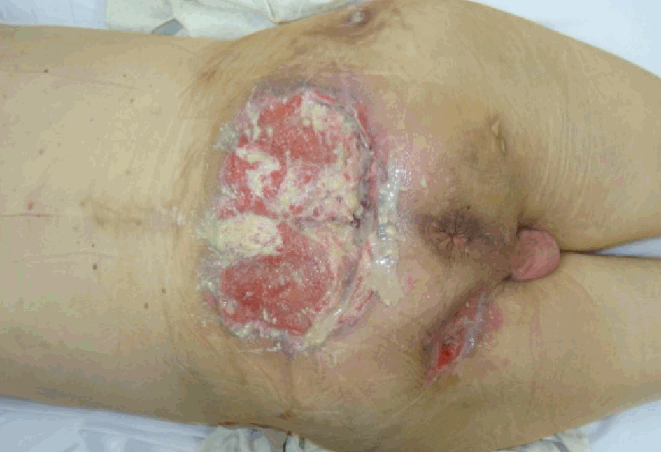
Marjolin’s ulcer on the sacral region involving the posterior surface of the sacral bone.
The only option for radical excision of the tumor was subtotal sacrectomy. Total sacral bone disarticulation between S1 and S2, ligation of the thecal sac, and removal of soft tissue and the residual gluteus muscles were performed. A posterolateral femur incision was made (Fig. 2). The popliteal vessels were ligated. Dissection was carried out through the posterolateral intermuscular thigh septum. The flap was harvested distally to proximally. The hamstring muscles were removed to avoid a bulk effect. We obtained a dually-structured flap, with a myo-fasciocutaneous portion (anterolateral side) and a fasciocutaneous portion (medial side) (Fig. 3). The anterolateral portion of the flap was vascularized by perforators from the superficial and deep femoral arteries, while the fasciocutaneous portion of the flap was supplied by perforators from the profunda femoris artery. The flap surface measured 50×38 cm (Fig. 4). The posteromedial component of the flap represented an extension of the flap that allowed the total flap surface to be increased; moreover, the fasciocutaneous component was vascularized by a well-known and secure pattern of perforators.
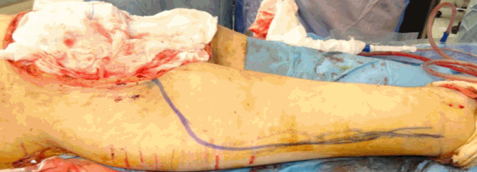
Posterolateral femur cutaneous incision planned for harvesting the subtotal extended thigh flap.

Subtotal extended thigh flap (A), containing a myo-fasciocutaneous anterolateral portion (solid line) and a fasciocutaneous medial portion (dashed line) (B).
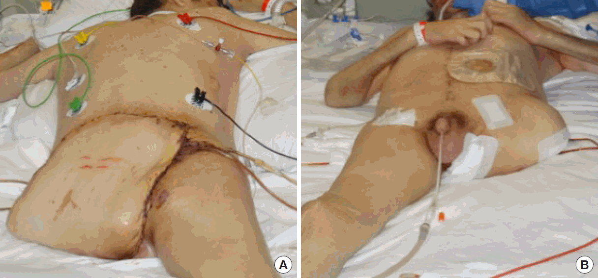
(A, B) Patient in the prone and supine positions during his 5th day in the intensive care unit.
This modification of the original flap enabled the flap surface to be enlarged, which could be useful in extreme cases of total or subtotal sacral resection.
Notes
No potential conflict of interest relevant to this article was reported.
Ethical approval
The study was approved by the Institutional Review Board of Campus Bio-Medico of Roma University and performed in accordance with the principles of the Declaration of Helsinki. Written informed consent was obtained. Written informed consent was obteined.
Patient consent
The patient provided written informed consent for the publication and the use of his images.
Author contribution
Conceptualization: all authors. Data curation: Tosi D, Marangi GF. Formal analysis: Delle Femmine PF. Funding acquisition: all authors. Methodology: Persichetti P. Project amministration: Persichetti P. Visualization: all authors. Writing original draft: Tosi D. Writing review and editing: Tosi D. Approval of final manuscript: all authors.