Peripheral osteoma of the infraorbital canal
Article information

Osteomas are benign osteogenic tumors arising from cancellous or compact bone [1]. They can be classified as central, peripheral, or extraskeletal. Peripheral osteomas arise from the periosteum as a pedunculated or sessile mass on the surface of the bone. They occur mainly in the paranasal sinuses, and most frequently in the frontal sinus [2]. This report presents a rare case of peripheral osteoma arising from the infraorbital canal [3].
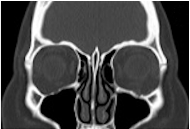
A 45-year-old woman was referred to our department with a complaint of left eye pain that had persisted for a year. A physical examination showed left proptosis, strabismus, and infraorbital nerve palsy. Her left intraocular pressure was elevated. Disturbance of eye abduction and depression were observed. Computed tomography (CT) revealed an 11 × 11-mm mass that protruded from the left infraorbital canal into the orbit, compressing the eyeball upward (Fig. 1).

Preoperative computed tomography (CT). CT revealed an 11×11-mm mass protruding into the orbit from the left infraorbital canal.
Surgery was performed with a subciliary approach under general anesthesia. A pedunculated tumor covered with mucoperiosteum was found on the floor of the orbit. The mass emerging from the infraorbital canal was located, and the infraorbital nerve was observed to be stretched on the surface of the tumor (Fig. 2A). The tumor was excised with a diamond bur, preserving the infraorbital nerve (Fig. 2B). The remaining defect in the orbital floor was about 3 mm. A histopathological examination revealed mature compact bone, confirming the diagnosis of a peripheral osteoma.

Intraoperative photographs. (A) A pedunculated tumor covered with mucoperiosteum was found in the floor of the orbit. The mass was continuous from the infraorbital canal (blue arrow), and the infraorbital nerve (yellow arrow) was observed to be stretched on the surface of the tumor. (B) The tumor was cut with a diamond bur, preserving the infraorbital nerve (yellow arrow).
Left infraorbital nerve palsy was observed immediately after surgery, and gradually improved. At a 6-month follow-up, the patient complained of minor hypoesthesia. Her extraocular movement fully recovered. Postoperative CT showed no evidence of recurrence (Fig. 3).
Postoperative 6-month image. There was no evidence of recurrence.
Notes
Conflict of interest
No potential conflict of interest relevant to this article was reported.
Ethical approval
The study was performed in accordance with the principles of the Declaration of Helsinki. Written informed consent was obtained.
Patient consent
The patient provided written informed consent for the publication and the use of her images.
Author contribution
Writing original draft, data collection, reference review: A Iwakami. Review, final editing: M Imanishi, H Asato. Approval of final manuscript: all authors.