Unusual presentations of philtrum of the lip
Article information
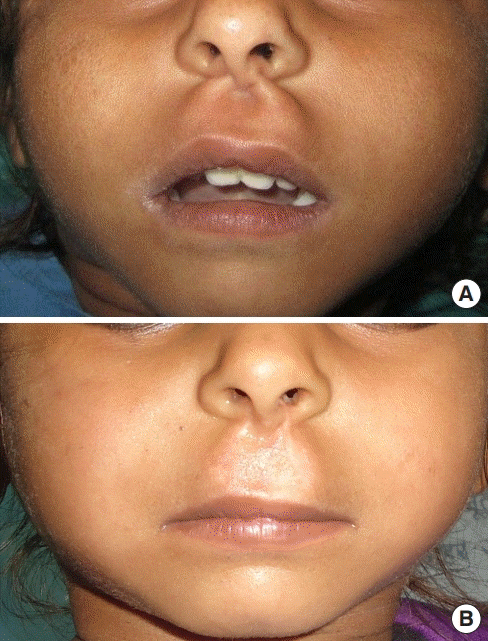
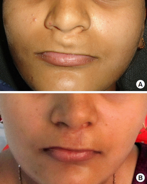
The philtrum is the most characteristic feature of the upper lip. Variations in its morphology can be seen amongst different individuals and races, but unusual presentations of the philtrum are relatively uncommon except in cases of orofacial defects, malformations, certain syndromes, trauma, and pathologies [1-3]. In our clinical practice, we encountered two cases involving an abnormal presentation of the philtrum, which were non-cleft, non-syndromic, non-traumatic, and non-pathological in nature. The first patient was a 6-year-old girl, who had thick ridges on both sides of the philtrum since birth (Fig. 1A). On gross examination, the height of the upper lip appeared relatively tall, the cupid’s bow was not distinct, and the philtrum was flat near the vermillion border. On palpation, the bands were firm and non-tender. We performed aesthetic correction of the bands with a satisfactory outcome (Fig. 1B). The second case was a 22-year-old woman, who had a philtral bulge at the base of the columella (Fig. 2A). This bulge was congenital and proportionately increased in size with her facial growth. She had no history of any infection or trauma. The philtral ridges, philtral depression, and cupid’s bow were indistinct. On palpation, the bulge was soft, non-tender, fixed to the overlying skin, and non-displaceable. On percussion, the fluid thrill test was negative. There was a small transverse scar near the base of the nose that was secondary to an incision made for drainage purposes when the child was 10 years old. We performed cosmetic reduction of the bulge and the results were acceptable (Fig. 2B).

A 6-year-old girl, who had thick ridges on both sides of the philtrum since birth. (A) Preoperative image. (B) Postoperative image.
Notes
Conflict of interest
No potential conflict of interest relevant to this article was reported.
Ethical approval
The study was approved by the Integrity Ethics Committee of CHL Hospital (approval No. CHL/DEN/JSC/JUN-2019/02) performed in accordance with the principles of the Declaration of Helsinki. Written informed consents were obtained.
Patient consent
The patients provided written informed consent for the publication and the use of their images.
Author contribution
Conceptualization, data curation, formal analysis, methodology, project administration, visualization: JS Chauhan, S Sharma. Writing - original draft, review, & editing: JS Chauhan, S Sharma. Approval of final manuscript: all authors.

