Septal deviation correction methods and surgical considerations in turbinoplasty
Article information

Abstract
Nasal septoplasty is often required to correct a cosmetic deformity, which is a common reason for patients to present to a plastic surgeon. If nasal septoplasty is insufficient, a residual deformity or nasal obstruction may remain after surgery. Even if the nasal septum is corrected to an appropriate position, nasal congestion could be exacerbated if the turbinate on the other side is not also corrected. Therefore, appropriate treatment is required based on the condition of the turbinates. Herein, we survey recent trends in treatment and review previous research papers on turbinoplasty procedures that can be performed alongside nasal septoplasty.
INTRODUCTION
Anatomically, the nasal septum consists of cartilage on the anterior aspect and bone on the posterior aspect. The anterior cartilaginous septum is particularly important in terms of functionality, as it is the most prevalent cause of nasal obstruction. The bony septum portion on the posterior aspect is relatively unimportant due to its larger cross-sectional area. However, posterior deviation sometimes occurs, with serious clinical symptoms. The causes of nasal septal deviation can be divided into intrinsic and extrinsic forces. Intrinsic forces can result in congenital or acquired septal cartilaginous abnormalities. Septal deviation due to extrinsic forces can occur as a result of scar contracture or asymmetric attachments of the osseocartilaginous skeleton. Nasal septal deviation should be corrected both because of nasal obstruction and because it can lead to nasal deviation [1].
Septal deviation may result in compensatory hypertrophy of the contralateral inferior turbinate. Even in the absence of preoperative airway obstruction, straightening the septum can cause the anterior airway to become narrowed if inferior turbinate hypertrophy is present. Therefore, several different management strategies can be used for inferior turbinate hypertrophy. We will discuss the basic techniques and points where surgeons need to be cautious, based on a review of various relevant studies.
TYPES OF NASAL SEPTAL DEVIATION
According to Guyuron et al. [1], nasal septal deviation can be divided into six categories, five of which may influence the external nose [2]. A localized septal deviation or spur has no effect on the direction of the external nose. The most common type of septal deviation is septal tilt, which is described as a septum that has no curve, but is tilted to one side of the nose anteriorly and to the opposite side posteriorly relative to the sagittal plane, with the maxillary crest remaining straight. In addition to anteroposterior septal tilt, cephalocaudal septal tilt can be found. The second category of septal deviation is C-shaped anteroposterior deviation. Unlike septal tilt, this deformity involves curvature of the cartilage. The third type is C-shaped cephalocaudal deformity. The fourth and fifth types of deformities are S-shaped septal deviations, which can be divided into anteroposterior and cephalocaudal. The last form of deviation is localized deviation or spurs. This type of deviation may accompany turbinate hypertrophy (Figs. 1-3).
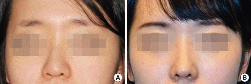
Septal tilt deviation
A patient with a septal tilt deviation preoperatively (A) and following straightening of the spreader graft and differential septal suture (B).
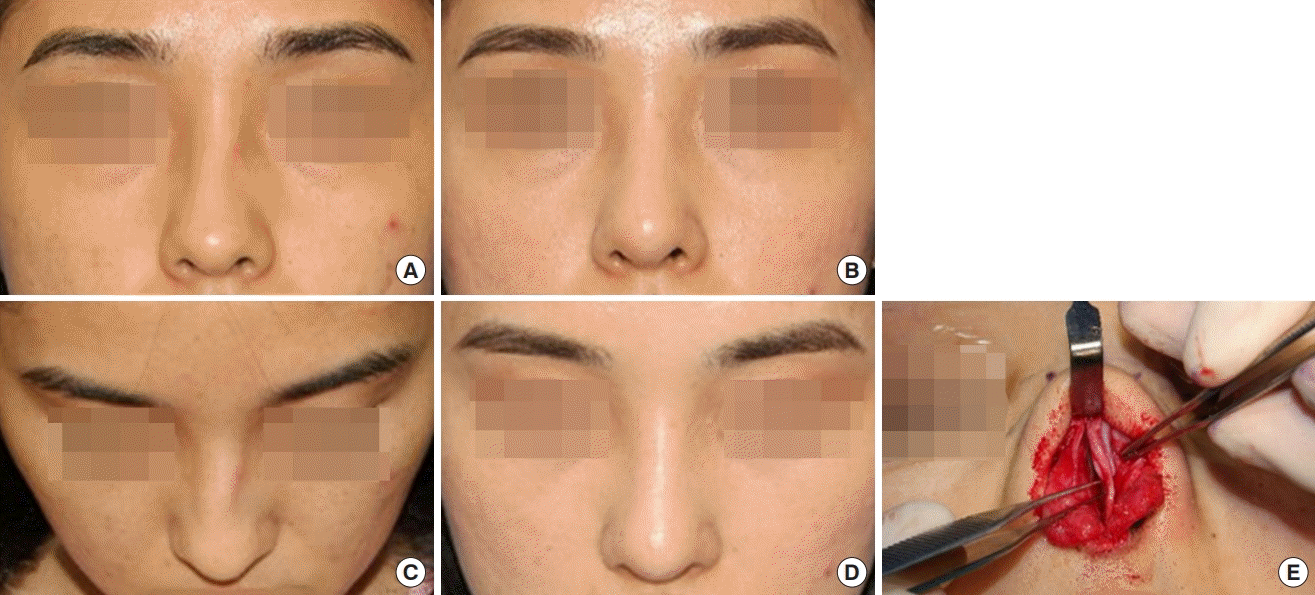
C-shaped septal deviation
A patient with a C-shaped septal deviation preoperatively (A, C) and following correction (B, D), intraoperative (E).
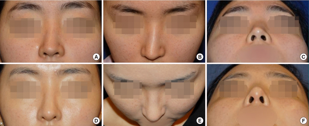
S-shaped septal deviation
A patient with a S-shaped septal deviation preoperatively (A-C) and following correction (D-F).
SURGICAL METHODS FOR NASAL SEPTAL DEVIATION
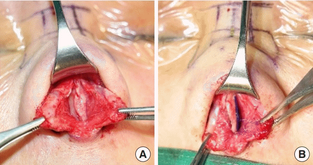
As nasal septal deviation occurs due to an intrinsic or extrinsic force, wide dissection of the mucoperichondrial flap is required. The septum and upper cartilage should be completely separated, and the anterior nasal spine (ANS) should be sufficiently dissected. If septum deviation is caused by an extrinsic force, the nasal septum may sometimes only be straightened with sufficient dissection. The caudal and dorsal L-struts should have a width of at least 10 mm; however, the precise width depends on the strength of the septal cartilage, and the struts may often need to be at least 15 mm wide to ensure long-term support [3]. The surgeon should remove the posterior-caudal portion of the cartilage to correct septal tilt. A quadrangular piece of cartilage should be completely separated from the maxillary crest and nasal spine, and partially mobilized with the perpendicular plate. The overlapping portion on the posterior side should be removed, and then the septum is repositioned with a figure-of-eight structure. Correction of a C-shaped deviation requires the posterior septum to be resected, leaving an L-strut. Scoring should also be performed on the concave side of the cartilage, completely freeing the septum and maxillary crest. The perpendicular plate is then partially released, along with a cephalic portion of cartilage. This type of deviation is corrected by adding a spreader graft anteriorly, at least on the concave side. An S-shaped deviation can be corrected by removing a posterior portion of cartilage, along with bilateral scoring on the concave side and resection of the protruding maxillary crest. A spreader graft can be used if necessary, and a batten graft may be added to straighten the remaining caudal septum (Fig. 4). In cases of localized septal deviation, removing the deviated portion can address most of what is needed for correction. In cases of high septal deviation, correction is not required to correct nasal obstruction, but it has been reported that limited septal correction and turbinate surgery can improve the symptom of nasal congestion [4]. However, if high septal deviation is accompanied by cosmetic asymmetry on the exterior, it needs to be rectified with procedures including a spreader graft, a differential septal suture, or a camouflage graft (Fig. 5).
C-shaped septal deviation
A patient with a C-shaped septal deviation preoperatively (A) and following correction (B), with a spreader graft and caudal batten graft (C, D).
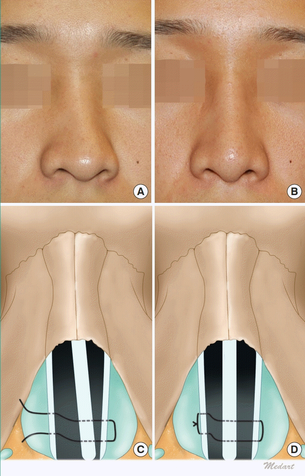
Septal tilt deviation
A patient with septal tilt deviation preopera-tively (A) and following straightening of a dif-ferential septal suture (B). The differential septal suture (C, D).
Furthermore, after correcting the cartilaginous portion of the nasal septum, if correction of the bony septum is insufficient, a bony spur may be removed using Takahashi forceps. If midline shifting of the bony septum is required, the microfracture technique may be performed, with extreme caution to prevent cribriform plate injury [5].
CORRECTION OF CAUDAL SEPTAL DEVIATION
Even after the posterior septum has been corrected, caudal septal deviation can still remain. Most such cases are accompanied by a vertical excess of the anterior septum. A completely separate caudal portion of the L-strut from the ANS and maxillary crest should be excised, the vertical excess should be removed, and then it should be repositioned on the ANS part. It can be fixed in the desired position through the periosteum using polydioxanone (PDS) 5-0 sutures or by drilling the ANS (Fig. 6) [3]. In some cases, it can be cut, leaving the base stump of the caudal septum, which is corrected by overlapping cartilage on the excess portion, and then reinforced with a batten graft. Ceran et al. [6] introduced a technique where the vertical excess of the caudal septum is removed, the gap between the ANS is reinforced, and PDS foil is used as a batten graft to straighten the caudal septum. However, this technique is not recommended, as the gap between the ANS and septum could weaken the long-term support of the caudal septum [7].
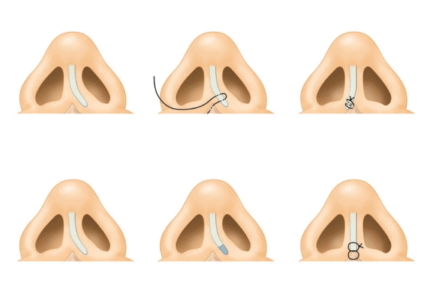
Caudal septal relocation suture
NASAL SEPTAL DEVIATION CORRECTION TECHNIQUES IN CASES WITH INSUFFICIENT NASAL SEPTAL CARTILAGE
In some cases, no nasal septal cartilage might be usable for primary nasal plastic surgery, and in other cases, a sufficient amount of cartilage cannot be obtained because the nasal septal cartilage is small. Although auricular or costal cartilage may be used to correct nasal septal deviation, PDS foil may be a good substitute. Some reports have confirmed the stability of PDS foil when used to correct complex cases of septal deviation, and found that it effectively maintained dorsal support over the course of long-term follow-up. PDS foil can also be useful as a batten graft in septoplasty, and it does not negatively influence graft take if it is placed on only one site of the cartilage (Fig. 7) [8]. The perpendicular plate of the ethmoid bone can also serve as a good source of material for grafting if the septal cartilage is unusable. According to the location of the deformation or deflection, it is possible to place a bone graft, which functions as an internal splint to reinforce the septal cartilage, at various positions along the L-strut [9,10].
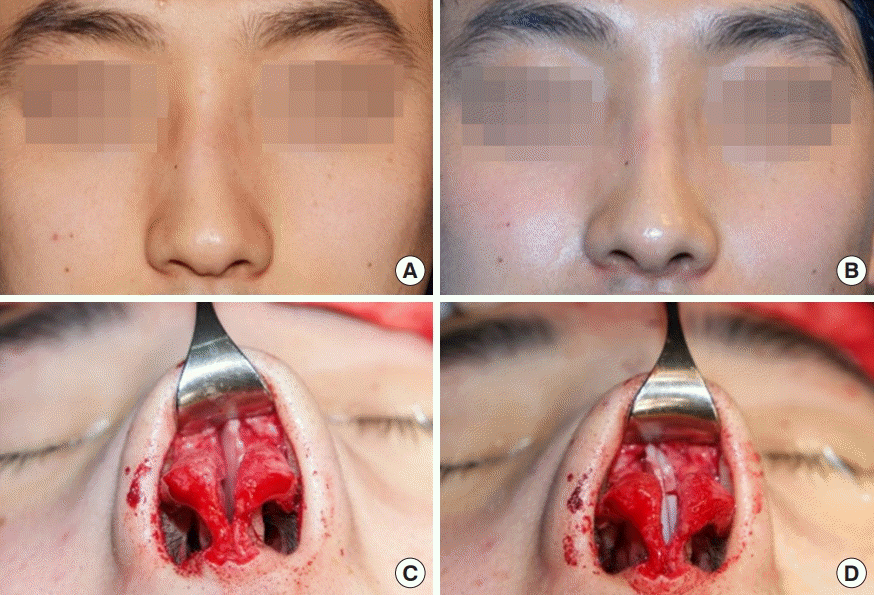
PDS foil used for caudal batten graft
(A) Caudal septal deviation. (B) Polydioxanone (PDS) foil for batten graft.
TURBINATE SURGERY FOR PATIENTS WITH NASAL SEPTAL DEVIATION
Septonasal deviations are often associated with airway problems. Septal deviation may result in compensatory hypertrophy of the contralateral inferior turbinate. Even in the absence of preoperative airway obstruction, straightening the septum may cause the anterior airway to become narrower if inferior turbinate hypertrophy is present. Complications of extensive surgical resection techniques are significant, with devastating outcomes including incurable rhinorrhea and empty nose syndrome. Out-fracture of the inferior turbinate was routinely performed in the past, but the turbinate position was found to rebound within months of surgery. The close microfracture technique, which is similar to the out-fracture technique, is a method of surgery with a lower likelihood of complications and minimal risk of recurrence. Unlike the out-fracture technique, the closed microfracture technique results in a comminuted fracture inside an intact mucosal sac, instead of a greenstick-type fracture with the potential to migrate back (Fig. 8). This technique is very simple. Using a long heavy Vienna nasal speculum, the inferior turbinate is microfractured through a closed approach. The turbinate is out-fractured in a juddering motion, proceeding from posterior to anterior while pushing the Vienna speculum laterally on the turbinate [11].
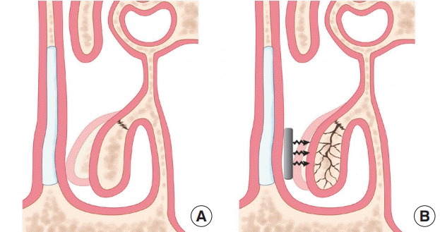
Microfracture of inferior turbinate
Traditional out-fracture (A) and closed microfracture for the treatment (B) of inferior turbinate hypertrophy
AUGMENTATION RHINOPLASTY FOR PATIENTS WITH NASAL SEPTAL DEVIATION
If augmentation rhinoplasty is performed simultaneously in patients suffering from nasal septal deviation, the septum and upper lateral cartilage should be reapproximated carefully after correcting the nasal septal deviation.
In particular, if the surgeon uses artificial materials such as a silicone implant, the perichondrium in the upper lateral cartilage should be sutured carefully to prevent penetration of the intranasal cavity. Moreover, the surgeon needs to exercise particular caution to prevent mucosal tearing. Nevertheless, if nasal osteotomy and septal deviation surgery are performed simultaneously due to septonasal deviation, augmentation rhinoplasty using autologous tissues such as a cartilage or dermis graft is recommended.
Notes
Conflict of interest
No potential conflict of interest relevant to this article was reported.
Patient consent
The patients provided written informed consent for the publication and the use of their images.