Dissecting cellulitis of the scalp: A diagnostic challenge
Article information

Dissecting cellulitis of the scalp (DCS), which is also referred to as Hoffman disease, is a rare chronic inflammatory disease of unknown etiology, commonly found on the vertex. Its clinical features are nodules, abscesses, and draining sinuses, often with scarring alopecia. Along with pilonidal cyst, hidradenitis suppurativa, and acne conglobate, DCS is part of the “follicular occlusion tetrad,” suggesting a common pathogenesis of deep follicular occlusion, followed by follicular rupture and subsequent follicular infection [1]. DCS is diagnosed based on clinicopathological features. Multiple treatment options, including antibiotics, retinoids, IMAGES corticosteroids, and tumor necrosis factor inhibitors, have been used with variable results [2]. However, surgical intervention is indicated in cases refractory to medical treatment [3]. Herein, we report a case of DCS to facilitate its recognition and proper management. An 18-year-old boy presented with multinodular lesions on the scalp (Fig. 1A). They were tender and fluctuant on palpation. No fluid was found on aspiration. He also had severe facial acne scars. Preoperative computed tomography showed lobulated soft tissue lesions (Fig. 1B).
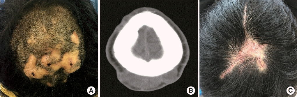
An 18-year-old boy with multinodular lesions on the scalp. (A) Preoperative photograph. (B) Preoperative axial computed tomography image showing lobulated soft tissue lesions. (C) Six-month postoperative photograph showing several tiny fistulas on the surgical scars.
Intraoperatively, the lesions were filled with chronic granulation tissue (Fig. 2). After removal of the lesions, the involved scalp was partially excised and closed. Tissue cultures and mycobacterial tests were negative. A histological examination showed granulation tissue formation with polymorphous inflammatory cells and entrapped keratin debris. The postoperative course was uneventful, except for several tiny fistulas on the surgical scars at the 6-month follow-up (Fig. 1C). The differential diagnosis of scalp nodules is extensive, and DCS often presents diagnostic challenges to surgeons due to its rarity. However, multinodular scalp lesions in patients with facial acne scars or lesions can be considered pathognomonic of DCS.
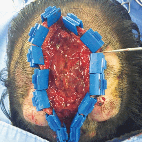
Intraoperative view showing the lesions filled with granulation tissue.
Notes
Conflict of interest
No potential conflict of interest relevant to this article was reported.
Ethical approval
The study was approved by the Institutional Review Board of Kangwon National University Hospital (IRB No. KNUH-2020-08-004) and performed in accordance with the principles of the Declaration of Helsinki. Written informed consent was obtained.
Patient consent
The patient provided written informed consent for the publication and the use of his images.
Author contribution
Conceptualization: SY Lee. Writing - original draft: KY Sung. Writing - review & editing, visualization: S Lee. Methodology, data curation: Y Jeong. Writing - review & editing: SY Lee. Approval of final manuscript: all authors.