Long V-Y advancement technique for large nipple reconstruction in Asian women
Article information

Abstract
Previously reported nipple-areolar complex reconstruction (NAR) methods involve multiple incisions and wide skin redraping, which increase retraction forces and heighten the risk of nipple-areolar complex (NAC) flattening. We introduce a NAR method using the long V-Y advancement technique that can overcome these disadvantages. A V-shaped flap is designed with the width of the flap base 4–5 mm larger than the diameter of the normal nipple. The flap length is designed to be at least 2.5 times its width. Dissection is performed to the top of the artificial dermal matrix or muscle layer. The nipple is constructed with the same projection as the contralateral side by folding the elevated flap. The tip of the elevated flap is apposed in the middle of the donor defect to minimize the deformity during donor site closure. A 3-point skin suture is applied to the upper third of the folded flap to mold its shape. Using this long V-Y advancement technique, we successfully decreased skin tension in NAC flaps and improved the maintenance of reconstructed nipple projection. The long V-Y advancement technique provides an easy, simple NAR method, effectively maintaining longer nipple projections and reducing breast deformities, especially in Asian women with relatively large nipples.
INTRODUCTION
Nipple-areolar complex reconstruction (NAR) is the final procedure during breast reconstruction following skin-sparing mastectomy, and various NAR methods using local flaps have been reported. However, these methods involve multiple incisions and wide skin redraping of donor sites, which increase retraction forces on the reconstructed nipple-areolar complex (NAC) and increase the risk of NAC flattening over time. After skinsparing mastectomy, breast skin is lacking and additional skin is needed for NAC reconstruction. Moreover, after tissue expansion, the expanded skin is relatively thin and provides insufficient support for the reconstructed NAC; thus, local flap-based NAR may deform the shape of the reconstructed breast. In particular, Asian women usually have smaller breasts with relatively large nipples, meaning that NAC flattening is of greater concern.
We introduce a NAR method using the long V-Y advancement technique that can overcome these disadvantages of local flapbased NAR.
IDEA
With the patient in the standing position, the new NAC is marked so that it is symmetrical with the normal side (Fig. 1).
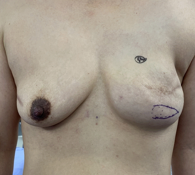
Preoperative image
The base of the V flap is set to the medial side, and the design is made to include the mastectomy scar in the lateral incision or to be parallel to the existing scar with slow tapering of the Vshaped tip. The width of the flap base is 4–5 mm larger than the diameter of the contralateral normal side and the flap length is designed to be at least 2.5 times its width. Dissection is performed to the top of the artificial dermal matrix or muscle layer.
The nipple is constructed with the same projection as the contralateral side by folding the elevated flap, and locations where the flap tip is to be sutured are marked. The tip of the elevated flap is properly apposed in the middle of the donor defect to minimize the deformity during donor site closure. A 3-point skin suture is applied to the upper third of the folded flap to mold its shape. Dog ears at both ends of the folded area are not trimmed to avoid compromising nipple circulation, and are subsequently molded by scar contracture during healing to produce a normal contour (Fig. 2). The flap dermal layer is then sutured with Vicryl 5-0 and the skin is closed with nylon 6-0 (Fig. 3).
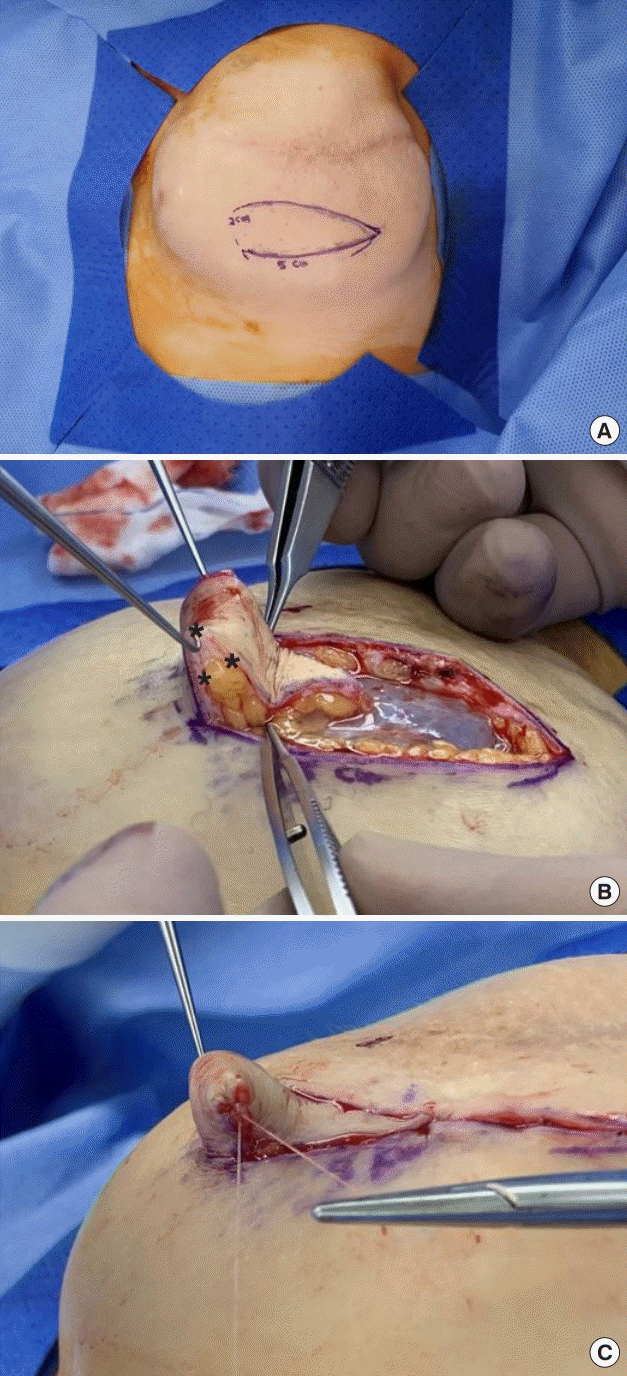
Surgical procedure
(A) The nipple base width is 4–5 mm larger than the diameter of the contralateral normal side and the length of flap is designed to be at least 2.5 times its width. The designed flap is elevated. The tip of the elevated flap is placed in the middle of the defect, rather than fixing it at the nipple base. (B) A 3-point skin suture is applied to the upper third area of the folded area to mold its shape (asterisks). (C) The cone-shaped deformities at both ends of the folded flap top are not trimmed to maintain nipple circulation.
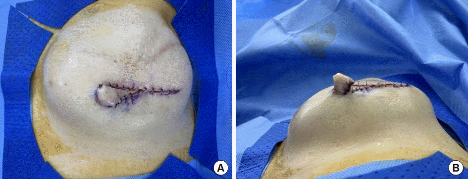
The reconstructed nipple
(A) Anterior view and (B) lateral view.
Antibiotic ointment and foam dressing are applied to protect the reconstructed nipple from compression.
Using this long V-Y advancement technique, we successfully decreased the skin tension in NAC flaps and improved the maintenance of reconstructed nipple projection. Fig. 4 presents the case of a 49-year-old patient who underwent right skin-sparing mastectomy and two-stage breast reconstruction (reconstruction using tissue expander and implants) with well-maintained nipple projection at 30 months postoperatively (initial projection, 9 mm; postoperative 30-month projection, 8 mm).
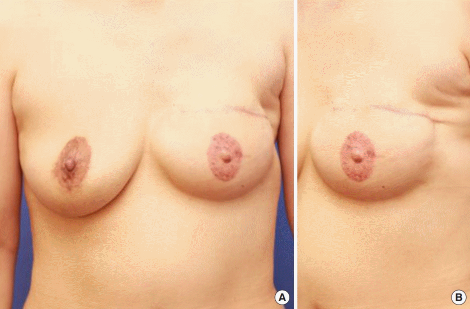
A case of nipple reconstruction
Medical photographs of a 49-year-old patient undergoing left skin-sparing mastectomy and two-stage breast reconstruction. Three months after twostage breast reconstruction, nipple reconstruction using the long V-Y advancement technique was performed. Anterior view (A) and anterolateral view (B) medical photographs taken at 6 months after nipple reconstruction, showing well-preserved long-term nipple projection and base width (initial projection, 4 mm; postoperative 6-month projection, 4 mm).
DISCUSSION
Many NAR methods, such as skate [1], star [1,2], C-V [3], and bell [1,4] flaps have been described, but all have inherent advantages and disadvantages. Furthermore, despite the wide variety of local flap designs, no consensus has been reached regarding which techniques are superior and when a certain technique should be employed. According to studies by Banducci et al. [2] and Bramhall et al. [5], despite advances in NAR methods, all reconstructed nipples show significant projection loss of around 25% to 50% at 2–3 months postoperatively and a 75% rate of unacceptable scarring. These outcomes can be explained by the absence of natural anatomic infrastructure, the centrifugal retraction forces below reconstructed nipples, and wound contracture [6].
Various methods have been introduced for the long-term maintenance of reconstructed nipple projection, using materials such as auricular cartilage [7], artificial dermal matrix [5], and subcutaneous inlay grafts [8]. However, these methods are not widely used due to multidirectional scars, foreign body reactions, additional donor site morbidity, prolonged operation times, and/or high cost.
The V-Y flap is a local advancement flap with a local pedicle that enables structure elongation and is commonly used for the closure of small to medium-sized cutaneous defects of the face [9]. We attempted to modify this technique and apply it to NAR.
The advantages of the long V-Y advancement technique are that the surgical method is straightforward, which greatly reduces the operation time, and that it enables the construction of large nipples. In addition, as a random pedicle flap, the base of the flap can be set in any direction, and it can therefore be applied to various existing scars or breast conditions. Furthermore, the technique makes it possible to leave only one Y-shaped scar, which reduces the scar burden and produces excellent cosmetic results. In addition, the reconstructed nipple receives sufficient blood supply, which decreases nipple flattening due to sufficient preservation of subdermal plexus, unlike other NAR methods (e.g., the skate or C-V flaps), which leave multiple scars around the nipple. Furthermore, it requires relatively little mobilization of the surrounding tissue for redraping and reduces peripheral breast deformation, which in turn reduces the retraction force on the reconstructed nipple. Thus, this technique minimizes NAC flattening.
Kroll [8] suggested that flap width is the most important factor for obtaining long-term projection, as increasing the width augments the flap blood supply and reduces fat necrosis. The long V-Y technique enables nipples to be sufficiently wide, allowing adequate blood flow. In C-V and skate flaps, which are made by collecting multiple flap lobules or tips in one place, blood supply is inevitably reduced at the tip area, potentially resulting in severe nipple flattening.
The advantages of a straightforward design, reduced scar burden, less tension, and sufficiency of blood supply ensure sufficient nipple projection and the safety of this devised method in patients, especially after second-stage reconstruction using a tissue expander with poor, thin, or tight skin and after autologous tissue-based reconstruction.
The application of this V-Y technique for NAR has been previously described by Riccio et al. [10]. However, these authors used this method for autologous tissue-based breast reconstruction and did not investigate the effect of applying his method to two-stage reconstruction with a tissue expander. Furthermore, the method they used involved placing the tip of the flap at the nipple base, which is the widest defect region, and thus, is subject to greatest tension during donor site closure and breast contour deformity. In addition, a small V-Y flap was used, which could lead to cosmetic dissatisfaction when applied directly to Asian women. Our proposed flap is designed to have a lengthto-base width ratio of more than 2:1, and the tip of the elevated flap is placed in the middle of the defect, rather than fixing it at the nipple base. This method of placement reduces tension during donor site closure, which can result in less breast tissue approximation, thereby reducing breast contour deformity and theoretically avoiding tension to the nipple base. In particular, for patients with thin, high-tension skin who undergo second stage reconstruction using a tissue expander, our proposed method provides an effective means of reconstructing the nipple that applies little tension to the surrounding breast tissue.
As mentioned above, trimming the flap margins may restrict distal circulation; thus, rather than trimming the cone deformity to produce a rounded shape, a 3-point skin suture was placed to form two small cones on each side of the folded flap. These cones were subsequently molded by scar contracture during the healing process.
We believe that this long V-Y advancement technique provides a simple means of NAR that effectively maintains longer nipple projection and reduces breast deformities, especially in Asian women with relatively large nipples. Further studies on nipple projection maintenance over the long-term are required to validate the described method.
Notes
Conflict of interest
No potential conflict of interest relevant to this article was reported.
Ethical approval
The study was approved by the Institutional Review Board of Kangbuk Samsung Medical Center (IRB No. KBSMC 2020-06-048) and performed in accordance with the principles of the Declaration of Helsinki. Written informed consent was obtained.
Patient consent
The patient provided written informed consent for the publication and the use of her images.
Author contribution
Conceptualization: J Kim. Data curation: N Jang, J Kim, HW Shin, SW Suk. Formal analysis: N Jang. Methodology: N Jang, J Kim. Project administration: N Jang. Writing - original draft: J Kim. Writing - review & editing: N Jang, HW Shin.