Application of nasal septal cartilage in a combined transorbital and transnasal approach for orbital wall reconstruction
Article information

The combination of the transorbital and endoscopic transnasal approach for the reconstruction of orbital wall fractures has been modified to be less invasive after the introduction of functional endoscopic sinus surgery. Artificial materials or iliac bone was used as graft material in past studies [1-3]. Instead, we use nasal septal cartilage as the graft material; herein, we describe its usefulness with a representative case. A 25-year-old woman had a blunt orbital injury.
Restriction of upward and downward movement of the left eyeball was accompanied by double vision. Computed tomography (CT) revealed herniated orbital tissue due to inferior and medial wall fractures (Fig. 1). Under general anesthesia, nasal septal cartilage was first harvested via endoscopy (Fig. 2). The orbital wall fractures were examined via an endoscopic transnasal and transconjunctival approach (Fig. 3). The herniated tissue was repositioned back into the orbit, and the bony defects were covered with the graft (Fig. 4). Postoperative eye movements were normal and double vision disappeared. An optimal cosmetic result was obtained without scarring or enophthalmos. Postoperative CT showed no evidence of recurrence. Nasal septal cartilage can be the first choice as a graft material for orbital wall reconstruction in the combined approach. First, it can be easily harvested to its maximum size under clear endoscopic vision at the start of the transnasal approach. Second, the morbidity is minimal and no additional incision is necessary. Third, it is a thin, flexible, and tractable autologous graft. However, it is necessary to use another graft material for a considerably large defect since the size of the nasal septal cartilage is limited.
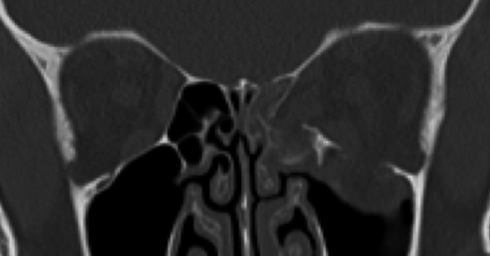
Preoperative computed tomography.
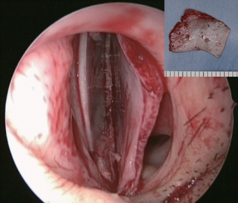
Nasal septal cartilage was harvested maximally with endoscopy.
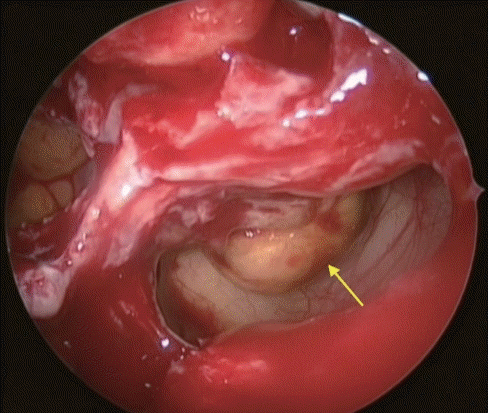
The fractures of the orbital walls and the herniated tissue (yellow arrow) were examined through the endoscopic transnasal approach.
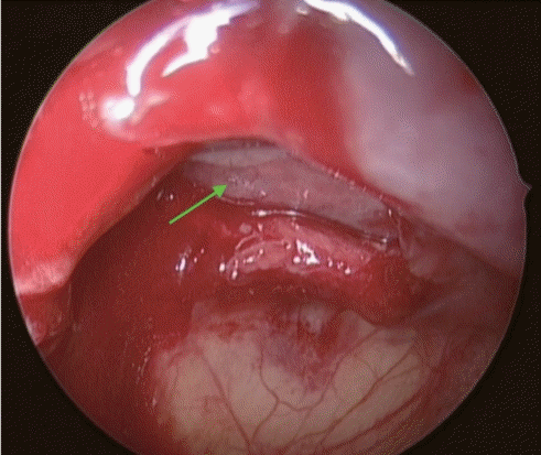
The herniated tissue was repositioned back into the orbit, and the bony defects were covered with the nasal septal cartilage (green arrow).
Notes
Conflict of interest
No potential conflict of interest relevant to this article was reported.
Ethical approval
The study was performed in accordance with the principles of the Declaration of Helsinki. Written informed consent was obtained.
Patient consent
The patient provided written informed consent for the publication and the use of her images.
Author contribution
Writing original draft, data collection, reference review: A Iwakami. Review: G Takada. Final editing: N Fukuda, H Asato. Approval of final manuscript: all authors.