Manual lymphatic drainage guided by real-time indocyanine green lymphography in breast cancer-related lymphedema
Article information

Lymphedema occurs when lymph vessels, collectors, or lymph nodes are damaged, and the physiological flow of lymph fluid is disrupted. This fluid accumulation is countered by an inflammatory response that worsens the lymphatic disruption and leads to a vicious cycle of fluid accumulation, inflammatory response, fat hypertrophy, and fibrosis.
The first line of treatment for lymphedema is complex decongestive therapy (CDT), which consists of a combination of different nonsurgical and noninvasive treatment approaches including bandaging, compression garments, exercise, skin care, and manual lymphatic drainage (MLD). The aim of CDT is to decrease the swelling of the affected limb to normal or near-to-normal size.
MLD is a safe procedure that may furnish additional benefits to compression bandaging in terms of swelling reduction [1,2]. It is a key component of CDT and should be performed by an experienced lymphedema therapist. MLD consists of soft skin stretching massages to help promote the movement of lymph fluid out of the affected Communication limb; it is focused on patent lymph vessels to help the flow of lymph fluid. This procedure stimulates alternative drainage pathways that prevent the stagnation of proteins in the interstitial space, thereby avoiding a vicious inflammatory cycle. Moreover, MLD has shown promising results in terms of preventing lymphedema after breast cancer surgery [3]. However, special care should be taken in patients with cardiovascular comorbidities or impairments of sensation, arterial circulation, or ability to eliminate the mobilized fluid, since MLD can move large amounts of fluid throughout the body. This category includes patients with diabetes, chemotherapy-induced sensory impairment in late-stage cancer, and peripheral neuropathy, among others. In addition, MLD may be less efficacious in patients with skin infections or wounds, motor deficits, or paralysis, which impair muscle activation in edematous body parts.
An accurate visualization of patent lymphatic vessels could guide the therapist during the treatment session and potentiate the effectiveness of MLD. In this regard, indocyanine green (ICG), a fluorescent cyanine dye that has a high rate of binding to plasma proteins and therefore remains within the blood and lymphatics, is ideal for visualizing superficial lymphatic vessels upon stimulation with fluorescent and laser light [4,5]. Hence, it is one of the most useful tools for the diagnosis and evaluation of lymphedema [5]. Although several studies have investigated ICG imaging for the diagnosis, staging, and surgical planning of patients with lymphedema, to the best of our knowledge, no report has yet been published on ICG lymphography-guided MLD (LG-MLD).
In 2018, the lead author (PC) started using ICG lymphography to guide the lymphatic therapist while performing lymphatic drainage. In our early experiences, LG-MLD has proven to be an objective way to assess lymphatic flow during the procedure. With this technique, the therapist can drain the lymph through viable lymphatic pathways and individualize the treatment, which may make the procedure more effective and efficient. It is particularly useful in patients with early-stage lymphedema, where a higher fluid component of the disease is present and lymphatic vessels are still patent and somewhat functional. It can also help patients to develop certain limb movements or exercises that can work better for moving the excess lymph fluid.
Our technique of performing lymphatic massage aims to open the lymphatic vessels to let the excess fluid drain back to the proximal lymph nodes. First, intradermal injections of 0.3 mL of ICG (Diagnogreen 0.25%; Daiichi Pharmaceutical, Tokyo, Japan) are performed in the second and fourth interdigital spaces in the hand of the affected extremity. An infrared camera is used to visualize the lymph channels. Figs. 1 and 2 depict a patient undergoing this procedure. Lymph flow can be seen in real time within minutes. The therapist uses hand movements to firmly but comfortably encourage the movement of the lymphatic fluid to a specific lymph node. Supplemental Video 1 shows the technique. ICG lymphography is only used on the initial visit to guide the first MLD session. Pictures (or video) and a sketch of the lymphatic network are uploaded into the patient’s chart to guide future sessions without the need for repeating ICG lymphography.
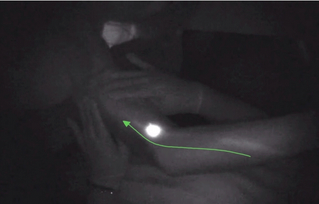
Lymphatic pathways are shown at the forearm and distal arm. The green line and arrow indicate the direction of the lymph fluid.
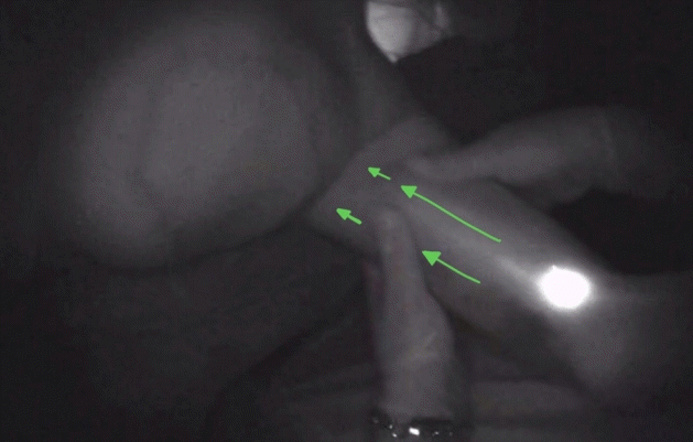
The lymphatic pathway is shown at the level of the arm. Green arrows indicate the direction of the lymph fluid. The therapist uses hand movements to firmly but comfortably drive the movement of the lymphatic fluid proximally.
In conclusion, ICG lymphography adds a real-time objective assessment of MLD and helps identify alternative pathways to tailor the massage in breast cancer-related lymphedema patients. The LG-MLD procedure is a valuable tool that may help improve the results of MLD. We are currently evaluating this procedure in a large series to further assess its potential long-term value for the treatment of extremity lymphedema.
Notes
Conflict of interest
No potential conflict of interest relevant to this article was reported.
Ethical approval
The study was performed in accordance with the principles of the Declaration of Helsinki. Written informed consent was obtained.
Patient consent
The patient provided written informed consent for the publication and the use of his images.
Author contribution
Conceptualization: P Ciudad. Methodology: P Ciudad, OJ Manrique, HC Chen, E Trignano. Project administration: P Ciudad, AJ Forte. Visualization: MT Huayllani. Writing - original draft: P Ciudad. Writing - review & editing: P Ciudad, SS Bustos.
Supplementary Material
Supplemental Video 1. Indocyanine green lymphography-guided manual lymphatic drainage. Supplemental data can be found at: https://doi.org/10.5999/aps.2020.01823.v001