Method to prevent cheek depression using an island sternocleidomastoid muscle flap with the middle pedicle as a feeding vessel in immediate reconstruction of the facial nerve with the sural nerve following resection of a parotid gland tumor
Article information

Abstract
Many surgeons have demonstrated the validity of sternocleidomastoid muscle flaps for the reconstruction of head and neck tumors. We present a case in which we used an island sternocleidomastoid muscle flap to reconstruct a cheek depression after excision of a malignant parotid tumor. A 44-year-old woman presented with a right malignant parotid tumor. We performed total resection of the parotid gland and facial nerve with the sural nerve and reconstructed the facial nerve and cheek depression with an island sternocleidomastoid muscle flap. The sternal head of the right sternocleidomastoid muscle was cut at the cranial and caudal segments to elevate it as an island flap. We used the superior thyroid artery as the sole pedicle for the island muscle flap. At 1 year and 3 months after the operation, the mimic muscles had gradually recovered and progressed without complications such as Frey syndrome, cervical motor dysfunction, or concave deformation of the neck and cheeks.
INTRODUCTION
Many reports have described tissue defect reconstruction after the excision of head and neck tumors using a sternocleidomastoid muscle or myocutaneous flap as a nerve bed [1-3]. An advantage of this method is that it provides additional blood flow for the transplanted nerve. We present a case in which we used an island sternocleidomastoid muscle flap filled with a middle pedicle for facial nerve reconstruction immediately after resection of a malignant parotid tumor. This case report highlights the importance of anatomical considerations. We report that our method of using an anatomically stable middle pedicle did not lead to an increased incidence of postoperative complications compared with reconstruction using a conventional pedicled sternocleidomastoid muscle flap. This method may be effective for preventing postoperative concerns such as mandibular bulge. This study was approved by the Ethics Committee of our institution (IRB No. 20-06-13) and was performed in accordance with the principles of the Declaration of Helsinki. The patient provided written consent for the publication and use of her images.
CASE
A 44-year-old woman presented with a tumor in the right parotid gland and facial nerve palsy. She scored 26 of 40 points according to the Yanagihara facial nerve grading system [4]. Based on the results of a fine needle aspiration biopsy, the patient was diagnosed with a malignant parotid tumor. Therefore, total resection of the parotid gland was performed, with an S-shaped incision in front of her right ear and facial nerve. After the malignancy was resected, the facial nerve and cheek depression were reconstructed using a sternocleidomastoid muscle flap that served as a nerve bed.
During resection of the malignant parotid tumor, the facial nerve was resected between the main trunk and the distal branch, which included the mandibular, buccal, zygomatic, and temporal branches. The sural nerve was used for facial nerve reconstruction. After harvesting, the sural nerve was divided into two parts, which were used for the nerve graft. In the upper part, the nerve graft was sutured with the facial nerve trunk and temporal branch in an end-to-end fashion. In the lower part, the proximal end of the nerve graft was sutured with the facial nerve trunk in an end-to-end fashion, and the four distal branches of the facial nerve were reconstructed using an end-to-side loop graft technique. The other end of the nerve graft was sutured with the ipsilateral hypoglossal nerve in an end-to-side fashion to supercharge the regenerating nerve fibers into the nerve graft. Subsequently, we elevated an island sternocleidomastoid muscle flap to serve as a nerve bed for the cheek depression after resection of the malignant parotid tumor.
In this patient, an island muscle flap that was nourished by the superior thyroid artery was raised to fill the defect area as follows: the sternum head was cut at the mastoid level to form the cranial side, and it was cut at the level of the upper two-thirds to form a caudal side matching the size of the defect.
The occipital artery could not be preserved because of concerns regarding possible limitations of muscle flap mobility. Finally, the island muscle flap was transferred without straddling the mandible (Figs. 1, 2).
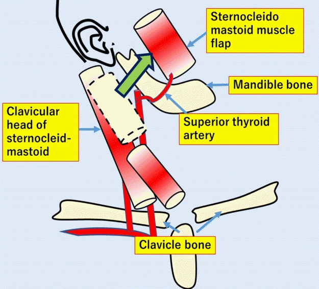
An island sternocleidomastoid muscle flap.
This schema outlines how we created the island sternocleidomastoid muscle flap.
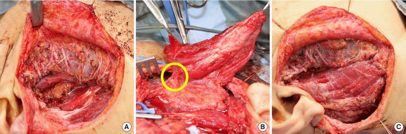
Operative photographs.
(A) Pre-reconstruction photograph. (B) The yellow circle identifies the point where the superior thyroid artery flowed into the sternocleidomastoid muscle. (C) The island sternocleidomastoid muscle flap was transferred to the parotid gland defect without tension.
One month after the operation, the patient received postoperative radiation therapy with a total dose of 60 Gy. At 1 year and 3 months after the operation, the facial muscles had gradually recovered and progressed without complications such as Frey syndrome, cervical motor dysfunction, or concave deformation of the neck and cheek (Fig. 3).
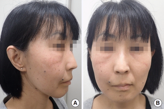
Postoperative results.
There was no swelling of the right mandible of the patient at 1 year and 3 months after the operation. (A) Profile view and (B) frontal view.
DISCUSSION
When filling a tissue defect after resection of a parotid tumor, preparing a nerve bed is an important goal. Motomura et al. [5] reported that using a muscle or myocutaneous flap as a nerve bed improves the blood flow of the tissue defect and is advantageous as an environment for the regeneration of the transplanted nerve. Moreover, covering the nerve graft with the flap prevents Frey syndrome, which can occur as a result of the auriculotemporal nerve intruding and filling the defect area of the parotid gland with thick tissue that prevents postoperative cheek depression. Furthermore, Tanaka et al. [6] found that for nerve reconstruction, performing nerve suturing after penetrating the nerve graft into the vastus lateralis muscle, even in patients who receive postoperative radiation therapy, can result in a better recovery of facial nerve palsy without delayed recovery. They also reported that a good postoperative course was obtained. However, their method, in which a conventional sternocleidomastoid myocutaneous flap was used as the superior pedicled sternocleidomastoid flap and the inferior pedicled sternocleidomastoid flap, the flap straddled the mandible, which resulted in the postoperative cosmetic problem of mandibular bulge. We were able to solve this problem by employing an island muscle flap supplied by the superior thyroid artery instead of a split sternocleidomastoid flap. An advantage of using the sternocleidomastoid muscle is that it is classified as type II according to the Mathes and Nahai classification due to its blood flow. Additionally, it is fed by one main artery and several minor arteries [7].
The blood supply of the sternocleidomastoid muscle is generally divided into three parts, including the upper, middle, and lower parts of the muscle body from its feeding arteries. The occipital artery is the main artery that flows into the upper part of the muscle, while the superior thyroid artery and the direct branch from the external carotid artery flow into the middle part and the superior scapular artery, the transverse carotid artery, the thyroid carotid artery, and the superficial carotid artery (sometimes several of them) flow into the lower part [8].
The results reported by Leclere et al. [9] revealed that, in the dissection of 15 cadavers, the blood supply to the sternocleidomastoid muscle was as follows: the upper one-third of the sternocleidomastoid muscle was 100% supplied by the occipital artery, while the middle one-third of the muscle was supplied by the superior thyroid artery or a direct branch of the external carotid artery or both, and other arteries were not observed. In contrast, in the lower part of the sternocleidomastoid muscle, the suprascapular, the transverse carotid, the carotid, and the superficial carotid arteries were mainly observed [10].
The patterns of the middle pedicle in the sternocleidomastoid muscle are simpler than those of the lower pedicles, which have complicated vessel patterns, and the superior thyroid artery or a direct branch from the external carotid artery is stably present in the middle pedicle. Thus, we showed that it was possible to raise a sternocleidomastoid muscle island flap with additional safety by using the middle pedicle instead of the lower pedicles. According to Leclere et al. [9], injection of latex into the inferior pedicle revealed that blood flow could be confirmed only up to the middle of the sternocleidomastoid muscle, but an injection into the middle pedicle confirmed blood flow up to the upper part of the sternocleidomastoid muscle in all cadavers.
In addition, a previous study reported that the occipital artery and the superior thyroid artery have anastomoses in the sternocleidomastoid muscle [9], and the blood flow from the middle pedicle reaches the upper part of the muscle. Therefore, a stable blood flow is ensured when creating a middle-pedicle sternocleidomastoid muscle island flap. This island muscle flap not only enhances mobility, but also ameliorates postoperative cosmetic concerns by avoiding having the muscle straddle the mandible.
To conclude, for immediate reconstruction of the facial nerve after resection of a parotid gland tumor, reconstruction was performed using a sternocleidomastoid muscle island flap with the middle pedicle as the feeding artery. With our method, which employs an anatomically stable middle pedicles, there is no increased incidence in postoperative complications compared with reconstruction using a conventional sternocleidomastoid flap. Thus, this method is considered to be advantageous in terms of the aesthetic surface after the operation.
Notes
Conflict of interest
No potential conflict of interest relevant to this article was reported.
Ethical approval
The study was approved by the Institutional Review Board of Yokohama Municipal Citizen’s Hospital (IRB No. 20-06-13) and performed in accordance with the principles of the Declaration of Helsinki. Written informed consent was obtained.
Patient consent
The patient provided written informed consent for the publication and the use of her images.
Author contribution
Conceptualization: H Sakuma. Methodology: A Shimono. Writing - original draft: N Matsuura. Writing - review & editing: all authors.