Independent finger-mounted device for vein visualization
Article information
Vein visualization during the harvest of vein grafts, flap elevation, and lymphovenous anastomosis formation can be made more efficient with the aid of a vein visualizer. We developed an independent finger-mounted device (IFMD) based on the concept of transillumination using pen-torches, as we have previously described [1]. This study was approved by our institutional review board. Transillumination works based on the differential light absorption of hemoglobin. Veins appear as silhouettes among adjacent tissues. Commercial transillumination devices are expensive, provide a limited visualization field, and do not allow effective illumination of surfaces with undulating contours due to their rigid framework [2]. Although near-infrared vein visualizers are effective, they are expensive and bulky [3]. The IFMD was developed with the aim of providing plastic surgeons with an inexpensive pocket-sized tool to assist in vein visualization.
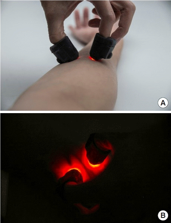
IFMD devices are mounted on the thumb and index finger and are swept across the region of interest to assess the superficial venous system (Fig. 1). Individual light sources constructed out of flexible silicone mounted on the fingers function comfortably as a natural extension of the user’s fingers. The device measures only 3×2 cm. The independent light sources are multidirectional and easily manipulated in three-dimensional space, catering to variations in contour and skin thickness. Light sources are directed to emerge beneath the veins, forming prominent silhouettes. Veins are identified in real time. The visualization field is increased by increasing the number of devices mounted (Fig. 2). The IFDM is cheap and easy to manufacture. It can be made easily accessible in resource-poor regions. Commercial vein visualizers are more expensive by up to a factor of 10 to 100.

The independent finger-mounted device. (A) Individual light sources conform to the contours of the forearm, channeling light for optimal vein visualization. (B) Vein over the forearm with branch points, visualized with two finger-mounted devices.

Additional application of the independent finger-mounted device. (A) Devices mounted on all digits to expand the field of visualization, an additional benefit of independent finger-mounted light sources. (B) Varicose veins visualized.
Notes
Conflict of interest
No potential conflict of interest relevant to this article was reported.
Ethical approval
The study was approved by the Institutional Review Board of National Healthcare Group, Singapore (IRB No. NHG ROAM DSRB 2018/01266) and performed in accordance with the principles of the Declaration of Helsinki.
Patient consent
The patients provided written informed consent for the publication and the use of their images.
Author contribution
Conceptualization, formal analysis, methodology, project administration, and visualization: EZ Cai, RS Hon, WT Chow, CC Yen, TC Lim. Data curation: EZ Cai, TC Lim. Funding acquisition: EZ Cai, TC Lim. Writing - original draft: EZ Cai, RS Hon, WT Chow, CC Yen, TC Lim. Writing - review & editing: EZ Cai, RS Hon, WT Chow, CC Yen, TC Lim.
