Linear scleroderma of the face treated by a superficial circumflex iliac artery perforator flap
Article information

Linear scleroderma, a subtype of localized scleroderma, is a rare disease that causes severe subcutaneous and deep tissue atrophy, which may result in a considerable psychological burden for patients [1].
Herein, we report the case of a 37-year-old female patient with linear scleroderma successfully treated with a superficial circumflex iliac artery perforator (SCIP) flap [2]. She had a linear brown plaque and marked atrophy of the skin and subcutaneous tissue of the left lower jaw that had lasted for 3 years (Fig. 1). The disease had been stable for more than 2 years. Preoperatively, the SCIP and superficial circumflex iliac vein (SCIV) were identified and marked with color Doppler ultrasonography. A 12 × 6 cm flap was designed, and an incision was made along the left inguinal ligament. Primary closure was performed at the donor site (Fig. 2). End-to-side and end-to-end anastomosis of the SCIP to the left facial artery and SCIV to the left facial vein, respectively, were performed. An adipose flap with only the monitoring flap was obtained by denuding. No relapse or complications were noted 3 months postoperatively (Fig. 3). Moreover, the concealed donor site was ideal for the young woman.
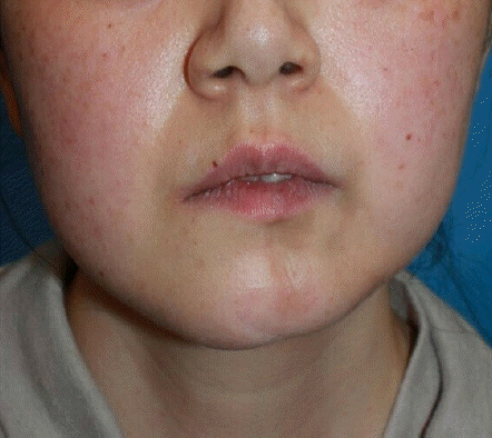
Clinical findings before the operation. At the patient’s first visit, there was a linear and sclerotic light brown plaque with marked atrophy on the left jaw.
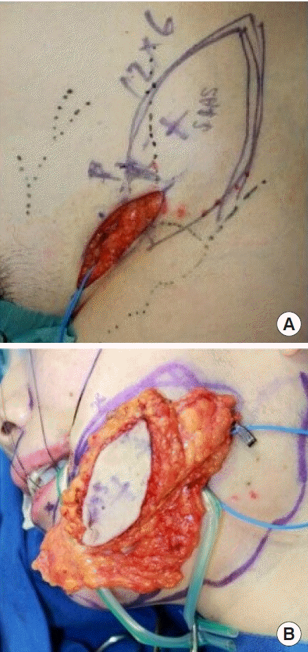
Intraoperative findings. (A) A 12×6 cm superficial circumflex iliac artery perforator (SCIP) flap was designed, and a surgical incision was made along the left inguinal ligament. (B) The SCIP flap was adjusted by minute defatting according to the degree of atrophy.
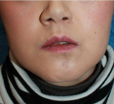
Clinical findings postoperatively. Three months after the operation, the contour of the patient’s face was bilaterally symmetrical.
Surgical treatment of facial atrophy caused by linear scleroderma requires tissue that is capable of withstanding wear and tear and is adjustable to various thicknesses [3].
For the complex reconstruction of facial tissue, a relatively noninvasive flap with a thickness that can be easily adjusted according to the location is ideal. The SCIP flap is likely to become an option for treating such cases.
Notes
Conflict of interest
No potential conflict of interest relevant to this article was reported.
Ethical approval
The study was performed in accordance with the principles of the Declaration of Helsinki. Written informed consent was obtained.
Patient consent
The patient provided written informed consent for the publication and the use of her images.
Author contribution
Conceptualization: H Mizuta. Writing - original draft: H Mizuta. Writing - review & editing: I Koshima. Approval of final manuscript: all authors.