Predicting lipoabdominoplasty complications with infrared thermography: a delta-R analysis
Article information
Abstract
The diagnosis of the main complications resulting from lipoabdominoplasty has not yet been standardized. Infrared thermal imaging has been used to assess possible complications, such as necrosis and changes in micro- and macro-circulation, based on perforator mapping techniques, among others. The objective of this study was to present two clinical cases involving thermal imaging monitoring of the healing process of lipoabdominoplasty in the immediate postoperative evaluation and its preliminary results. Infrared thermography was performed 24 hours after the operation and on postoperative days 5, 25, and 27. In clinical case 1, it was found that the delta-R (∆TR)–defined as the difference in minimum temperature between the highest and lowest points in the SA3 region (caution suction area) following the classification established by Matarasso–was 0.4°C at 24 hours after surgery and decreased to 0.1°C on a postoperative day 5. There were no complications in this case. In contrast, in clinical case 2, the ∆TR was 1.7°C at 24 hours after surgery (upon hospital discharge) and remained high, at 2.2°C, on postoperative day 5. A higher ∆TR was found in the second patient, who developed necrosis of the surgical wound. The ∆TR thermal index may be a new tool for predicting possible complications, complementing the clinical evaluation and therapeutic decision-making.
INTRODUCTION
Abdominoplasty is one of the most widely performed procedures in plastic surgery in the world, accounting for 15.9% of cases of plastic surgery performed in Brazil, according to the Brazilian Society of Plastic Surgery [1]. In recent decades, the techniques for abdominal contour surgery have undergone modifications and enhancements that have yielded improvements in aesthetic and functional results, in addition to reducing the incidence of complications [2]. In 2001, Saldanha et al. [3] described for the first time lipoabdominoplasty using the principles of liposuction and abdominoplasty with preservation of the abdominal flap and upper and lower muscle-cutaneous perforating vessels, with less impairment of the nervous and lymphatic systems. This technique reduced the incidence of complications, such as seroma, epitheliolysis, dehiscence, and necrosis, from 60%, 3.8%, 5.1%, and 4% in classic abdominoplasty to 0.4%, 0.2%, 0.4%, and 0.2% in lipoabdominoplasty, respectively. These complications seem to be essentially related to the anatomical limits of the surgical procedure. Not respecting these limits through excessive detachment of the abdominal flap creates more dead space and tension in the sutures. The result of lipoabdominoplasty with limited infraumbilical undermining is preservation of the important abdominal perforators of the deep inferior epigastric artery and a significant reduction in ischemic flap problems postoperatively [2-5].
Currently, the diagnosis of the main complications after plastic surgery on the abdomen has not been standardized; diagnoses are often still made by clinical examinations, through the visualization of bulging, palpation, and puncture. Ultrasonography is an imaging option that can assist in the diagnosis of seromas and bruises, followed by magnetic resonance imaging in some difficult cases [4]. Other complementary exams such as Doppler ultrasonography, photoplethysmography, venous occlusion plethysmography, 133Xe clearance, transcutaneous pO2, the heat wash-out technique, and laser Doppler imaging help to assess the vascular system and the tissue reperfusion status in plastic surgery. However, these methods are not done on a routine basis because they have some disadvantages such as a lack of portability, standardization and calibration problems, radiation exposure, the need for trained operators, and the high cost and delays in the acquisition and review of imaging studies [6].
In recent years, increasing interest has emerged in the use of infrared thermal imaging in plastic surgery for the functional assessment of microcirculation and mapping of perforators with high-definition images and sensitivity [7-9]. Infrared thermal imaging is a painless, totally safe, non-contact examination that does not emit radiation; it measures and maps the temperature distribution of the skin surface with a precision of tenths of degrees [6-9]. Infrared thermography also enables assessments of the sympathetic vasomotor response of the skin, inflammation, tissue perfusion, blood flow and the metabolic activity of tissues [6,10]. The more hyper-radiant (i.e., hot) the tissue is, the greater its activity, which can suggest dysfunctions and skin complications, as well as a more hypo-radiant (i.e., colder) condition. More attentive care in the immediate postoperative period, especially monitoring the inflammatory process inherent in tissue healing, is directly linked to a reduction in complications after lipoabdominoplasty [4,5].
The aim of this study was to present two clinical cases involving thermal imaging monitoring of the healing process of lipoabdominoplasty in the immediate postoperative evaluation and its preliminary results, with the goal of ascertaining whether thermal imaging has the potential to be used for the predictive evaluation of possible complications, in addition to complementing the clinical evaluation and therapeutic decision-making. Clinical case 1 presented a postoperative course without complications, whereas clinical case 2 presented a postoperative course with necrosis of the surgical wound. Based on these cases, thermal imaging appears to be promising for predicting possible complications after lipoabdominoplasty.
IDEA
Cases
Case 1
A 50-year-old female patient with no associated diseases, who was a non-smoker, underwent lipoabdominoplasty. In the immediate postoperative period, she wore a low-compression mesh (8 mmHg) and an anatomical flexible polyurethane abdominal splint (R-Slim). Infrared thermography was performed 24 hours after the operation and on days 5, 25, and 27 postoperatively.
Case 2
A 46-year-old female patient with no associated chronic diseases, who was a non-smoker, underwent lipoabdominoplasty and mastopexy with a prosthesis. She wore a low-compression (8 mmHg) mesh in the immediate postoperative period. Infrared thermography was performed 24 hours after the operation and on days 5, 25, and 27 postoperatively.
Thermal images
Verbal informed consent was obtained from the participants. The patients were instructed not to take a hot bath within 2 hours before the exam, not to use any topical substances on the skin, and to fast for at least 3 hours before the exam. During the 10-hour period before the exam, they were instructed not to consume any stimulants (such as coffee, alcohol, or cigarettes). Infrared thermography was performed 24 hours after the operation and on postoperative days 5, 25, and 27. The thermal images were captured using T530SC double-spectroscopy infraredvisual camera (Termocam; FLIR Systems, Wilsonville, OR, USA), with an image resolution of 320 × 240 pixels, thermal sensitivity of 0.05°C, and cutaneous emissivity set at 0.98. The exam was carried out at a controlled room temperature of 23°C ± 1°C, with a relative humidity of less than 60% and minimum air convection of 0.2 m/s. The subjects were allowed to rest comfortably without any clothes covering the abdomen for 15 minutes, and the images were captured at a distance of approximately 50 cm at right angles.
The images were analyzed by software developed by one of the authors (MLB). The “muscle-2” color scale was used in the double spectroscopy mode (VisionFy; Thermofy, São Paulo, Brazil).
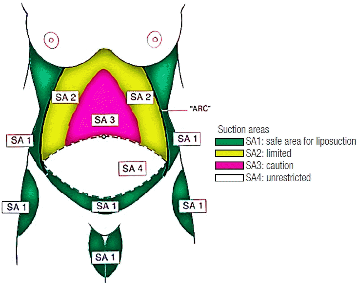
For the analysis of thermal images, the Huger zones, as reviewed by Matarasso (Fig. 1) [11], were used as regions of interest (ROIs) for reference; these zones take into account the vascular anatomy of the anterior abdominal wall and the blood supply of that region.
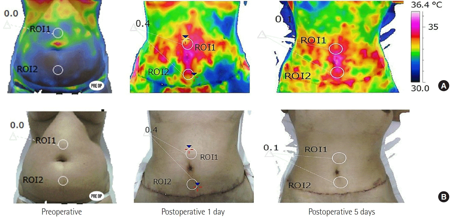
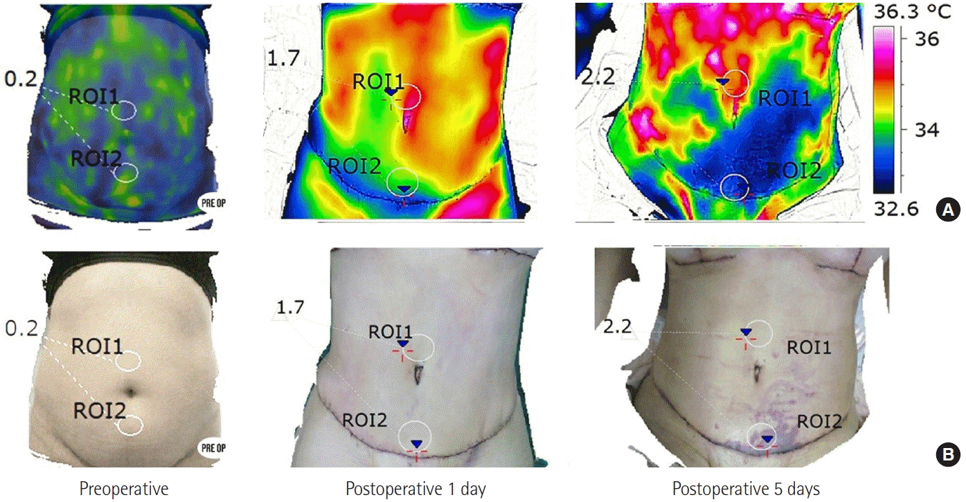
The SA3 zone (caution) was chosen, and two ROIs were determined in this area: ROI1 at the highest point (supraumbilical) and ROI2 at the lowest point above the suture, due to the random and axial blood supply to the abdomen. To assess the risk of complications due to ischemia or tissue inflammation, we calculated the values of the delta-R (∆TR), defined as the difference between the minimum ROI1 temperature and the minimum ROI2 temperature (Figs. 2-4).

Good postoperative course of lipoabdominoplasty: preoperative and postoperative images. (A) Multispectral thermal images and (B) corresponding photographs. The highest (ROI1) and lowest (ROI2) points in the SA3 region (caution suction area) were selected to calculate the differences in minimum temperatures. In images on the postoperative day 1, ∆TR=0.4°C. In images on the postoperative day 5, ∆TR=0.1°C. ROI, region of interest.

Poor postoperative course of lipoabdominoplasty: preoperative and postoperative images. (A) Multispectral thermal images and (B) corresponding photography. The highest (ROI1) and lowest (ROI2) points in the SA3 region (caution suction area) were selected to calculate the differences in minimum temperatures. In images on the postoperative day 1, ∆TR=1.7°C. In images on the postoperative day 5, ∆TR=2.2°C. ROI, region of interest.
Results
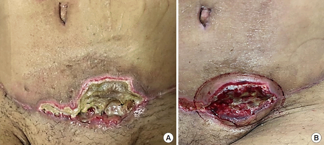
In clinical case 1, the ∆TR was 0.4°C at 24 hours after surgery and decreased to 0.1°C on a postoperative day 5 (Fig. 2). In contrast, in clinical case 2, the ∆TR was 1.7°C at 24 hours after surgery (at the time of hospital discharge) and remained high, at 2.2°C, on postoperative day 5 (Fig. 3). A higher ∆TR was found in the patient who developed necrosis. To illustrate the course of case 2, new photographs of the patient were taken 25 and 27 days after surgery (Fig. 4).
DISCUSSION
Infrared images enabled an assessment of the postoperative prognosis by identifying and measuring the difference in the skin temperature between predetermined risk regions. In view of the limited clinical evaluation, infrared thermography can assist in the early detection of unexpected changes and possible complications, such as epidermolysis, in the first postoperative days. Thermography allowed us to check the tissue reperfusion status and predict flap necrosis, making it possible to take measures to minimize tissue damage early.
An anatomical and physiological assessment and knowledge of the vascular system and the inflammatory process are essential for the planning and postoperative care of lipoabdominoplasty. The diagnosis of cutaneous necrosis after abdominoplasty can be presented visually in a simple way, as signs of self-limited epidermolysis and small dehiscence until the occurrence of extensive necrosis with loss of substance in the deep plane [11].
Temperature is an inflammatory sign that can be accurately measured by infrared thermography before visual clinical changes [6]. In the immediate postoperative period, the expected physiological effect is an increase in blood flow with a consequent increase in temperature, as was observed in clinical case 1 (Fig. 2), in this sense, the use of thermal imaging makes it possible to detect hypo-radiation (a decrease in temperature), as observed in clinical case 2 (Fig. 3) and, consequently, allows an assessment of decreased perfusion that is much more sensitive than a visual or tactile clinical evaluation. To perceive a thermal difference with the back of the hands, a difference greater than 2°C is necessary between the area of interest and the underlying normal skin; with an infrared sensor, the sensitivity is greater than 0.05°C (that is, 40 times the capacity of humans); furthermore, this method makes it possible to represent and delimit the risk area using imaging [12].
There is always the possibility of reduced blood flow in the region surrounding the surgical incision, with consequent impairment of oxygenation and delivery of nutrients to the wound; therefore, we suggest calculating the ∆TR in the SA3 region of Matarasso [11] using the minimum temperatures of ROIs 1 and 2, because these areas correspond to the location of the main angiosomes and perforating vessels responsible for the nutrition of the flap (critical area) [3,9,11].
The initial results obtained suggest that there is a clinical correlation of necrosis as a complication and ∆TR measured by infrared thermography. There was greater thermal asymmetry in the area of caution in case 2, in which epidermolysis was diagnosed on postoperative day 5 and skin necrosis eventually developed. It should also be kept in mind that the presence of seroma and hematoma usually presents as hypo-radiance on thermal images, with a more diffuse distribution, that is, not limited to the anatomical area described by Matarasso and the angiosomes of that region. Thus, greater differences in temperature (∆TR) may suggest, in addition to the impairment of microcirculation and metabolic tissue activity, more serious complications and a worse prognosis.
It is recommended to avoid excessive liposuction, closure under tension, and the use of very tight compressive meshes to reduce the risk of ischemia [2,4,5]. Likewise, the formation of edema and stasis of liquids in the extravascular spaces, which can lead to the plugging of small local vessels, should be avoided [13]. It is important to note that the pattern of compression in the postoperative period of these two cases was different. The patient in case 1 used, in addition to the low-compression mesh, a semi-rigid anatomical polyurethane splint, while the patient in case 2 used only a compressive mesh. It is worth mentioning that the meshes fold in the midline of the abdomen, regardless of the type of compressive mesh used; therefore, these folds can exert greater pressure on the midline of the abdomen and, following the Matarasso areas, can decrease blood flow in the caution area [11]. The authors believe that the bending of the midline caused by the brace in the patient in case 2 may have been a negative factor affecting the course of the case, as shown by the thermal images and photographs.
It is thought that infrared thermal imaging can be used to predict possible complications, in addition to complementing the clinical evaluation and therapeutic decision-making when performed correctly and methodically. The decrease in complications after lipoabdominoplasty is due to the growing concern with immediate postoperative care, combined with monitoring of the inflammatory process inherent to tissue healing; thus, interest has emerged in the application of infrared thermography to monitor the physiological processes that impact the temperature of the tissues [2,3,5].
Studies on the use of thermography in the field of plastic surgery have shown promising results. Most of them verified the use of infrared thermography to assess the location and quality of perforating vessels in the preoperative, intraoperative, and postoperative period, and concluded that the thermographic examination helps in planning surgery and achieving more efficient results with a reduction in complications [7-9]. The results presented herein should be confirmed by studies with a greater number of cases to verify the effectiveness and sensitivity of infrared thermography for predicting the prognosis of lipoabdominoplasty, as well as in the healing process after other plastic surgery procedures, in order to avoid further complications. The importance of establishing references for normal and pathological values of ∆TR in early monitoring of the healing process in plastic surgery is also emphasized for these values to serve as safer parameters for early decision-making.
The potential of infrared imaging can be verified by the early diagnosis of the evolution and complications in the postoperative period of lipoabdominoplasty. Furthermore, this technique also allowed objective monitoring of treatment when using the proposed methodology. The authors observed in the postoperative period of lipoabdominoplasty that hypo-radiating areas were related to microcirculatory complications, such as hematomas and seromas. Thus, greater temperature differences in the caution area may suggest necrosis even before clinical visual and tactile signs, such as epidermolysis.
Since after any surgery, there is a possibility of reduced blood flow in the specific region of the surgical incision, with consequent impairment of oxygenation and nutrient delivery to the wound, it is recommended to calculate ∆TR in the SA3 region of Matarasso [11] using the minimum temperatures of ROIs 1 and 2, the areas containing the main angiosomes and perforating vessels responsible for the nutrition of the flap (critical area). Based on the findings of this study, the proposed technique of thermal monitoring is promising as a new tool predicting possible complications and complementing the clinical evaluation and therapeutic decision-making.
Notes
Conflict of interest
No potential conflict of interest relevant to this article was reported.
Ethical approval
The study was approved by the Institutional Review Board of Hospital das Clínicas, Faculty of Medicine, University of São Paulo (IRB No. 891/19) and performed in accordance with the principles of the Declaration of Helsinki. Verbal informed consent was obtained from the participants.
Patient consent
The patients provided written informed consent for the publication and the use of their images.
Author contribution
Conceptualization: P Rodrigues Resende, ML Brioschi. Data curation: P Rodrigues Resende, F De Meneck, E Borba Neves, M Jacobsen Teixeira. Formal analysis: ML Brioschi, F De Meneck, E Borba Neves. Methodology: ML Brioschi, F De Meneck, E Borba Neves. Project administration: M Jacobsen Teixeira. Visualization: P Rodrigues Resende, E Borba Neves, ML Brioschi. Writing - original draft: P Rodrigues Resende, F De Meneck, E Borba Neves. Writing - review & editing: ML Brioschi, E Borba Neves, M Jacobsen Teixeira.


