Delayed ventral hernia repair after skin graft closure of the open abdomen: the use of tumescence for safe skin graft elevation
Article information

Recent advances in temporary abdominal closure techniques have allowed safe management of the open abdomen after damage control laparotomies. The eventual goal of managing such patients is closure of the fascial defect as soon as clinically feasible. When fascial closure is not possible, skin-only coverage over healthy granulation and planned delayed ventral hernia repair is the only option. If adequate skin is available, it can be primarily closed over the fascial defect. Otherwise, split-thickness skin grafting can be performed once adequate granulation tissue forms over the bowel [1]. A planned delayed ventral hernia repair is subsequently performed once the skin graft matures over the bowel. In most instances, a pseudomesentery develops between bowel and graft, which becomes evident with a “pinch test.” This provides a safe plane from which the skin graft is elevated off the bowel before definitive hernia repair is performed.
Nonetheless, the interface between bowel and skin graft may not be very well-defined in some cases, and the margin of error may become dangerously narrow during dissecting a plane between the skin graft and bowel.
We describe the use of tumescence for hydro-dissection and improving the safety margin when elevating skin grafts from the underlying bowel. This case is a 70-year-old male with a background history of diabetes mellitus and ischemic heart disease. He sustained polytraumatic injuries following a road traffic accident in 2018. His list of injuries included multiple rib fractures and a severe splenic injury complicated by abdominal compartment syndrome. An emergency laparotomy and splenectomy were performed followed by temporary abdominal closure. This was followed by several subsequent relook laparotomies and negative pressure wound therapy dressings. After sufficient granulation had formed, split-thickness skin grafting was performed to the central abdominal wound a month after the index surgery.
A large ventral hernia subsequently developed with an abdominal wall defect size of 35 × 32 cm (Fig. 1). He returned for definitive repair of the abdominal wall defect approximately 2 years later. The mature skin graft was excised safely and with ease after hydro-dissection with tumescent infiltration.
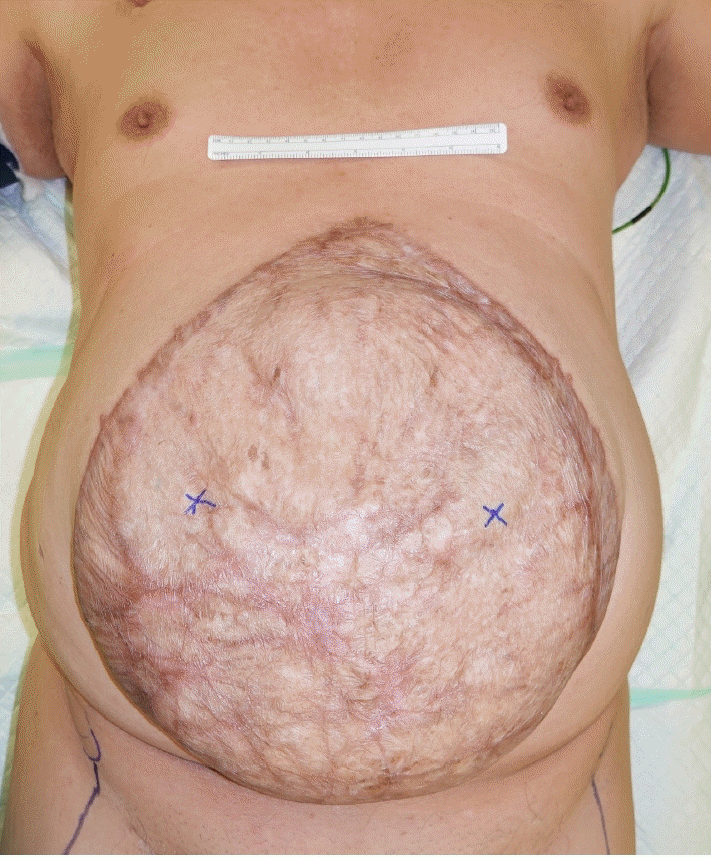
An abdominal wall defect size of 35×32 cm.
Normal saline solution was used for tumescent infiltration in the subcutaneous plane with a blunt liposuction cannula and a 50 mL syringe. Stab incisions of 2–3 mm were made with a 15-blade at entry points around the borders of the skin graft. To avoid penetrating into deeper tissues, the tip of the cannula is angled at a slight upward angle during insertion, and always visualized just under the skin (Fig. 2). The skin is also tented up prior to infiltration, during which superficial spreading of tumescent solution confirms the correct tissue plane. Elevation of the skin graft is performed easily and safely with sharp dissection using scissors (Fig. 3). Anterior component separation was performed, and a biological mesh was used to bridge the remnant abdominal wall defect.
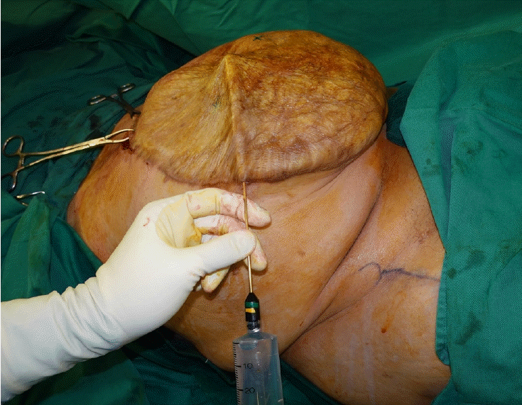
Insertion of a cannula with its tip angled upwards and always visualized during infiltration to avoid inadvertent bowel injury.
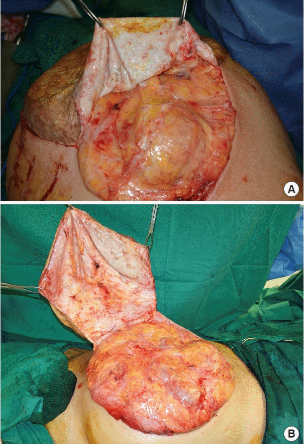
Safe elevation of skin graft from underlying abdominal viscera, with a clearly visualized plane of dissection (A, B).
The concept of “damage-control” surgery was born from the recognition that critically ill patients are more likely to die from the vicious triad of coagulopathy, acidosis, and hypothermia, than from the completion of surgical repairs [2]. This led to the emphasis on abbreviated surgery and prompt return to intensive care and aggressive resuscitation. Hence, patients are often left with an open abdomen, with a plan to return to the operating room for definitive repair of injuries. Various temporary abdominal closure techniques exist, and allow maintenance of sterility in-between operations without compromising intra-abdominal pressures [3].
Definitive fascial closure during the initial hospitalization may not be possible in patients who have undergone multiple reoperations, have ongoing intra-abdominal infection, visceral edema, or loss of fascial substance. In such circumstances, planned ventral hernia is the only option. Skin coverage options in such cases include skin-only closure, or a split-thickness skin graft once adequate granulation tissue forms over the bowel. The latter option can be further supplemented by an absorbable mesh (e.g., Polyglactin 910 mesh) to prevent evisceration [1]. A ventral hernia ensues and often times, the patient is planned for elective repair. The skin graft matures over 6 to 12 months, forming a stable connective tissue interface between the bowel and skin. Once this occurs, the adhesions mature to a filmy stage, allowing the skin graft to separate from the underlying tissue [4,5]. This is determined clinically by a positive pinch test, when the skin graft can be pinched without grabbing the underlying viscera. Nonetheless, a clear and safe plane may not always be evident between the skin graft and underlying bowel, resulting in a very narrow margin of error during graft elevation. Furthermore, despite allowing adequate time for skin graft maturation, a positive pinch test may not be uniformly apparent throughout the grafted field, resulting in unpredictable areas of closely adherent bowel. Notwithstanding meticulous surgical techniques, inadvertent bowel injury is a possibility during skin graft dissection [5]. This may lead to infectious complications and prolong the healing process.
Tumescence involves local subcutaneous infiltration of a solution to produce a swelling under the surgical site. Solutions may include various combinations of adrenaline for its hemostatic properties, normal saline, and a local anesthetic agent. It is commonly used in a number of plastic surgery procedures including liposuction, hair transplantation, cervicofacial rhytidectomy, mammoplasty, abdominoplasty and flap surgery. Apart from its content-dependent advantages in reducing intraoperative bleeding and providing anesthesia, tumescence facilitates surgical dissection by hydro-dissection.
This is especially important in cases such as ours, where tumescent infiltration helps create a clear plane of separation between skin graft and the underlying bowel through hydro-dissection. Hydro-dissection also loosens scar tissue and adhesions in that plane and increases the thickness of tissue through which separation occurs, further facilitating dissection while reducing the risk of shearing and bowel wall injury during skin traction. This improves the margin for error during dissection through adhesions, reduces the amount of sharp dissection required, accelerates large areas of skin elevation, and improves the overall safety of such a procedure. Furthermore, using a blunt liposuction cannula carries a low risk of bowel injury, especially when used in the above-mentioned manner.
To date, there are no fail-safe methods to reduce bowel injury during skin graft elevation in delayed ventral hernia repairs. Tumescence is a well-established technique used in a variety of plastic surgery procedures. Its novel application as described above will allow the surgeon to elevate the skin graft safely and expediently, and ultimately alleviate the risk of bowel injury.
Notes
Conflict of interest
No potential conflict of interest relevant to this article was reported.
Ethical approval
The study was performed in accordance with the principles of the Declaration of Helsinki. Written informed consent was obtained.
Patient consent
The patient provided written informed consent for the publication and the use of his images.
Author contribution
Conceptualization: HW Ng. Formal analysis: K Koh. Methodology: HW Ng. Project administration: HW Ng. Writing - original draft: K Koh. Writing - review & editing: K Koh.