Squamous Cell Carcinoma Arising from an Epidermal Inclusion Cyst
Article information
Epidermal inclusion cysts are commonly encountered benign cystic lesions that can occur anywhere in the body. However, malignant changes are rare, and only a few cases of malignant change have previously been reported [12].
Anton-Badiola et al. [2], based on a total of 13 reported cases, found that the mean age at presentation of squamous cell carcinoma arising from an epidermal inclusion cyst was 43.2 years. They also observed that it occurred more frequently in men, that the head and neck area was the most commonly affected region, and that the mean diameter was 5.7 cm.
Although a specific triggering factor for this malignant transformation has not yet been established, a well-known risk factor for malignancy is chronic irritation of the skin. Several possible factors, such as sun-related actinic damage and human papilloma virus skin infection, have been suggested to be triggering factors [2345]; however, more research regarding the direct influence of these factors on malignancy is needed.
A 62-year-old male presented with a cyst on his left cheek that had grown rapidly in size over the course of three months (Fig. 1). Physical examination revealed a mildly hard, fixed, bulging, and painless mass measuring 2 cm in diameter.
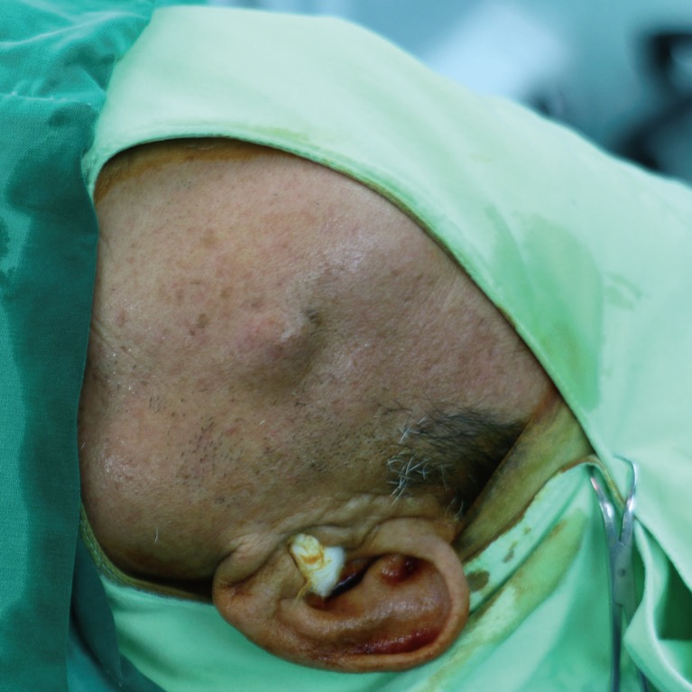
A 62-year-old male who presented with a 2×2-cm soft bulging mass on the left cheek.
At first, we thought this was likely to be a simple cystic mass. Thus, only a straightforward ultrasonography study was performed, leading to the biopsy of a benign-appearing palpable mass lesion in the cheek, performed under local anesthesia. The permanent biopsy was performed immediately by a pathologist. Contrary to expectations, the pathologic finding was well-differentiated squamous cell carcinoma arising from an epidermal inclusion cyst.
Gross examination showed an elliptical patch of skin measuring 2.5×2.5 cm, with an attached nodular lesion measuring 2.0 cm in diameter with an irregular margin. Upon dissection, the cut surface revealed a cystic mass containing yellowish keratin material.
Microscopically, malignant cells were found to have infiltrated the interior of an epidermal cyst, comprising 30% of its total size. Multiple neoplastic cells with large nuclei were found next to an epidermal inclusion cyst (Fig. 2). These pleomorphic cells appeared to arise from the overlying squamous epithelium of the cyst (Fig. 3). These cells showed an irregular patterned contour and obvious nucleoli with abnormal mitosis (Fig. 4). All of these features indicated a lesion of well-differentiated squamous cell carcinoma arising from an epidermal inclusion cyst.
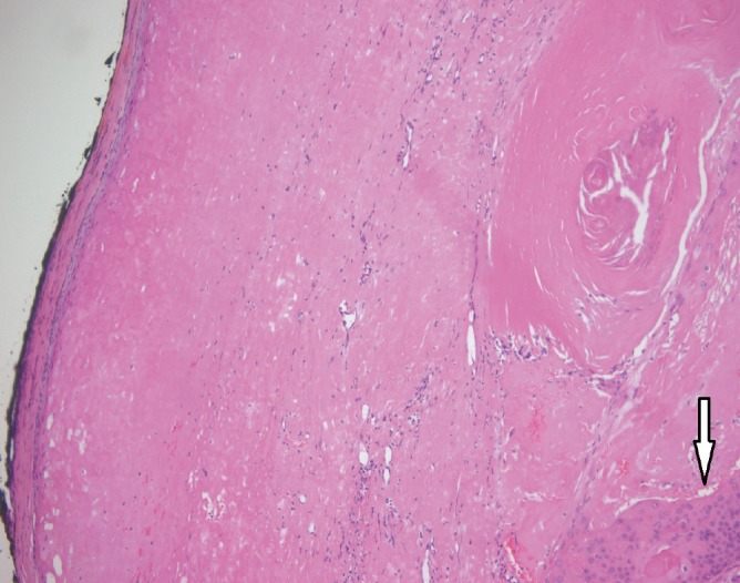
Neoplastic cells with large nuclei (white arrow) were found under the epidermal inclusion cyst containing keratin (H&E, ×100).
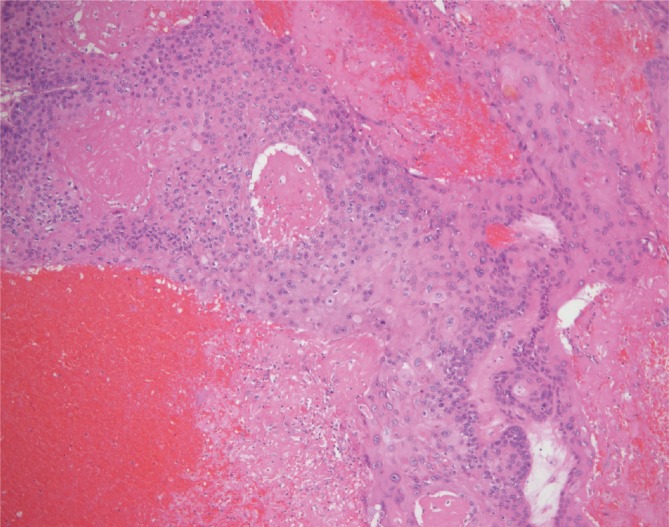
The tumor cells had large nuclei and showed pleomorphism of each cell (H&E, ×100).
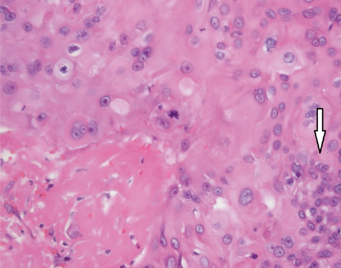
Normal epithelial cells have a regular margin with a circular shape. Malignant cells have an irregular polygonal shape (white arrow) (H&E, ×400).
Further evaluation was then performed. Contrast-enhanced computed tomography showed a heterogeneous and irregular margin in the subcutaneous fat layer with no cervical lymph node metastasis. A Tc-99m whole body bone scan showed no bone metastasis. Seven days later, reoperation was performed to extend the surgical margin, and total excision with a 1-cm margin was performed under local anesthesia. Free surgical resection margins were observed on the frozen biopsy. After the excision, the skin and soft tissue defect measured 4.0×4.0 cm and the defect was covered by a bilateral V-Y advancement flap. No clinical evidence of recurrence was observed more than one year postoperatively.
In our case, the patient had a mass that rapidly grew over the course of three months. We suggest that the rapid growth of the mass was an element of tumoral pathogenesis, causing the benign epithelium to undergo abnormal dysplastic change.
Epidermal inclusion cysts and squamous cell carcinoma are commonly encountered skin lesions in practice. The malignant transformation of these cysts is rare, and few cases showing such malignant changes have been previously reported.
The etiology of malignant transformation in epidermal inclusion cysts remains uncertain. Chronic irritation of the lesion has been frequently suggested as a triggering factor.
Due to the infrequency of malignant changes, not all epidermal inclusion cysts are routinely excised, and not all excised cysts are sent to pathologists for accurate examination.
In summary, despite the rarity of malignant transformations of epidermal inclusion cysts, we suggest that malignant changes should be suspected in cases showing rapid growth, ulceration, or frequent recurrence. Moreover, complete excision with pathological examination should be performed in all cases of epidermal inclusion cysts to avoid misdiagnosis.
Notes
No potential conflict of interest relevant to this article was reported.