Non-Invasive Imaging of Preoperative Mapping of Superficial Veins in Free Flap Breast Reconstruction
Article information
Vascular anatomy is imperative in reconstructive surgery to ensure perfusion and accurate flap design, but venous anatomy can be difficult to determine preoperatively. Anatomic landmarks can be used to locate structures; however there can be considerable variation in anatomy and location of veins. Techniques such as radiographic imaging, Doppler probes, and ultrasonography have been used to assess veins. However, radiographic imaging incurs a cost and radiation exposure. Doppler probes are easy to use but are more useful for arterial anatomy. Ultrasonography is limited by cost, time, and accessibility.
Several vein visualization devices, including the AccuVein (Avant Medical, Cold Spring Harbor, NY, USA), have been previously reported as an adjuvant to locate veins [1] and to aid intravenous cannulation. These devices were helpful to visualize veins, especially in difficult patients (extremes of age, obese, dark-skinned) [234]. The AccuVein is a portable device (size, 5×6×20 cm; weight, 275 g) that emits a red laser at 642 nm and an infrared laser at 785 nm. Hemoglobin preferentially absorbs infrared light so the skin the skin illuminated a red color and veins are silhouetted as dark outlines (Fig. 1). The color schemes can also be inverted to display veins as red silhouettes on the skin.
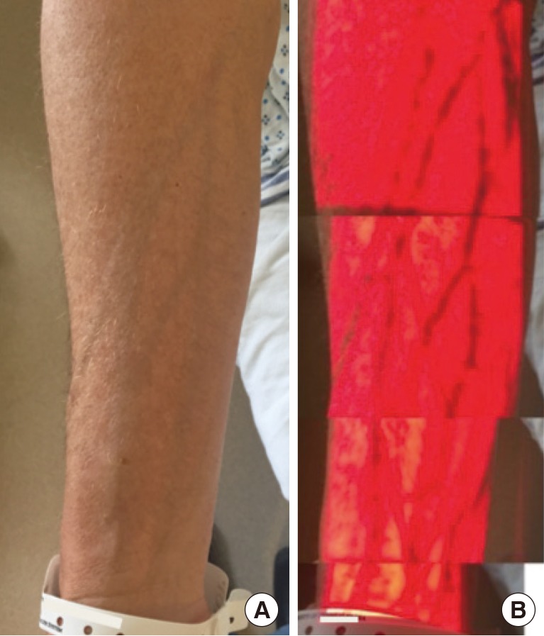
(A, B) Hand-held vein visualization device, (AccuVein) uses a red laser and infrared laser to illuminate basilic and cephalic venous vasculature for the forearm.
The AccuVein device was used in our study for abdominal-based free flap breast reconstruction to aid in locating abdominal veins and to evaluate candidacy for superficial inferior epigastric artery (SIEA) perforator flaps. To the best of our knowledge the use of this device in breast reconstruction has not been reported.
The current practice at our institution for planned free flap breast reconstruction involves intraoperative decision-making to determine flap type [5]. Routine preoperative radiographic imaging is not obtained. After making the inferior incision on the abdomen the SIEA is carefully identified. If the SIEA is absent, injured, or of insufficient caliber (<1.5 mm) then an SIEA flap is not used and the deep inferior epigastric system is assessed next. Medial and lateral row perforators are identified and a decision is then made to use either a deep inferior epigastric artery perforator flap or a muscle-sparing free transverse rectus abdominis musculocutaneous flap [5].
In our study the AccuVein was used for preoperative venous mapping of the superficial inferior epigastric veins (SIEV), superficial circumflex iliac veins, and to help guide dissection and flap design of the inferior skin paddle incision (Fig. 2). If an adequate SIEV was visualized preoperatively, the inferior skin incision was occasionally redesigned and made more inferior to ensure inclusion of the vascular pedicle before it arborizes.
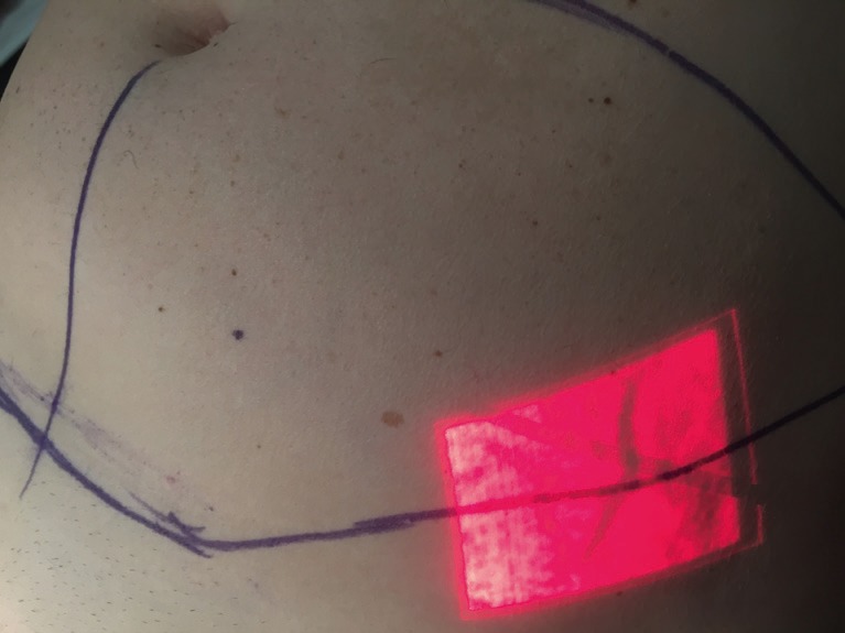
Preoperative visualization of left superficial epigastric vein before breast reconstruction.
The AccuVein was used on ten patients. In eight patients at least one SIEV was identified preoperatively in all patients and there was 100% positive and negative correlation of preoperative markings with intraoperative exploration (Fig. 3), and all veins were able to be identified intraoperatively. In the other two patients had abdominal striae that interfered with venous imaging and abdominal veins were not able to be visualized. In two patients preoperative vein mapping changed the flap design: the inferior incision was lowered to explore the vein before it bifurcates to include a larger caliber vessel in the flap for flap drainage or when patients were possible SIEA flap candidates. There were no AccuVein related complications and no false negatives that led to adverse flap planning. There were also no other perioperative complications or microvascular complications or flap losses for all ten patients and patients were on average, discharged on postoperative day 4.1 (range, 4–5).
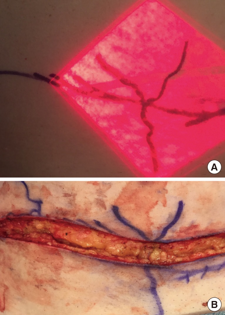
(A, B) Preoperative and intraoperative photographs of superficial epigastric vein during the donor site dissection of abdonimal-based free flap breast reconstruction using right hemi-abdomen.
The AccuVein is a non-invasive, portable, fast, and easy to use device to assess veins in real time. The disadvantages of this device are that it only produces a two dimensional image and the vein is displayed based on vessel content and not the actual vessel caliber. Although there were no false positives in our study, we recommend direct visualization of vessel caliber before any surgical decisions are made. The AccuVein can provide preoperative information about venous anatomy that can be useful in aiding dissecting veins and flap design.
The AccuVein is not necessary to successfully perform free flap breast reconstruction but we believe the AccuVein is an effective adjuvant that can help aid flap design and expedite dissection. The SIEV is a useful lifeboat that can be used as venous outflow for abdominal-based flaps and also used as a vein graft. Anatomic landmarks exist for most critical structures, and the SIEV is also consistently found midway between the anterior superior iliac spine and the midline. But anatomy can be variable and the AccuVein can expedite dissection and flap design.
Preoperative vein mapping can be useful for those starting to perform SIEA perforator breast free flap reconstructions. Preoperative venous mapping can help guide flap design and speed dissection of the SIEV and SIEA vena comitans. Radiographic imaging provide much more information than can AccuVein, but also costs more, is invasive if contrast-enhanced imaging is used, may require radiation exposure, and needs time to schedule, perform, and review images. On the other hand, the AccuVein device is often already available in many hospitals in the nursing units and is a simple, cheap, noninvasive, and quick method for preoperative mapping of superficial veins.
Notes
No potential conflict of interest relevant to this article was reported.