Sepsis Leading to Mortality after Augmentation Rhinoplasty with a Septal Extension Graft and Fat Grafting
Article information
Infection is the most serious complication after rhinoplasty, with rates reported to range from 2.6% to 5.3% [1]. In that study, most infections were found to be localized and improved after removal of the implant and appropriate antibiotic therapy. Although a case report has described staphylococcal spinal osteomyelitis after septoplasty as a consequence of bacteremia [2], no case of sepsis resulting in death after rhinoplasty has been reported. We report the case of a male patient who expired due to infection after rhinoplasty with a septal extension graft, silicone implant, and fat grafting, and then review and discuss the relevant literature.
A 27-year-old previously healthy male patient underwent blepharoplasty, augmentation rhinoplasty with a silicone implant, a septal extension graft with a strut graft, and the grafting of retroseptal fat that was harvested during blepharoplasty at the local hospital onto the nasal tip for tip projection. Erythema occurred in the nasal tip and columella on the first postoperative day, and ceftriaxone was therefore prescribed. On the second postoperative day, the patient complained of a general itching sensation caused by an allergic reaction, and the antibiotic regimen was then changed to a combination of ciprofloxacin and amikacin. Nevertheless, the erythema of the nasal tip worsened and wound disruption occurred (Fig. 1). On the tenth postoperative day, the silicone implant and septal extension graft were removed in our hospital (Fig. 2). Intraoperatively, extensive tissue necrosis and signs of infection were observed in the septal mucosa (Fig. 3).
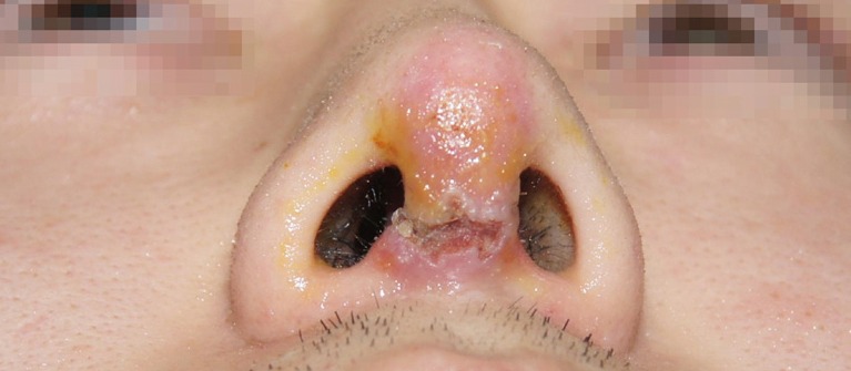
Preoperative view. Erythema on the nasal tip and columella is shown. The incision wound was disrupted.
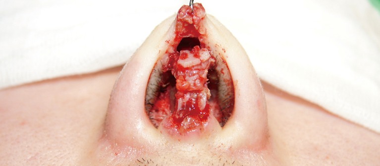
Intraoperative view. The silicone implant was removed. The onlay graft and columellar strut graft are shown.
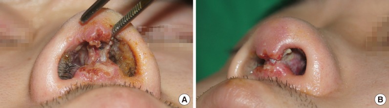
Intraoperative view. (A, B) The septal extension graft was removed. Extensive tissue necrosis and signs of infection were observed in the septal mucosa. This picture was taken on the day of the operation in our hospital and ten days after surgery at a local clinic.
For infection control, 400 mg of ciprofloxacin was administered twice a day and massive irrigation was performed. However, fever occurred and symptoms related to sepsis appeared on the third day (Table 1). Therefore, 1,500 mg of amoxicillin was prescribed as a broad-spectrum antibiotic pending the results of the susceptibility test. Five days after the debridement, the wound culture from the grafted fat was identified as methicillin-resistant Staphylococcus epidermidis. We changed the antibiotic prescription to 1 g of vancomycin twice a day based on the results of the susceptibility test. Nonetheless, the patient's general condition worsened. We transferred the patient to a tertiary care university hospital, but he eventually expired three days later.
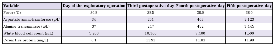
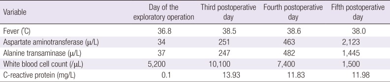
Changes in the laboratory results of the patient over the course of the infection
The nasal septum adjoins the brain, and a case of S. pneumoniae meningitis after septoplasty as a result of this anatomical feature has been reported [3]. However, our patient's cerebrospinal fluid was normal and no signs of meningitis, such as nuchal rigidity or mental status change, were noted.
In the present case, the cultured organism was methicillin-resistant S. epidermidis, a type of so-called superbacteria. However, the patient showed no response to antibiotics, making it difficult to ascertain that S. epidermidis was the primary cause of sepsis. Administering broad-spectrum antibiotic therapy within one hour after the diagnosis of severe sepsis is recommended, and the antibiotic regimen should be reassessed every 24 hours for de-escalation. Reassessment of the antibiotic therapy needs to continue after the susceptibility test results are reported, and the antibiotic therapy must be changed when septic conditions are escalated. The choice of antibiotics depends on the suspected site of the infection, the setting in which the infection developed, the patient's medical history, and local microbial-susceptibility patterns [4]. The blood culture, the results of which were only available after the patient expired, showed no growth. This may have been a false negative due to antibiotic coverage. The exact cause of sepsis was not determined by autopsy, which found no evidence of underlying disease related to impaired immunity, including leukemia and lymphoma.
This was a rare case in a young man, although infections can easily spread from the septal mucosa, which has a rich blood supply, to the brain and to the entire body. Therefore, aggressive treatment of the affected area is the best strategy to prevent sepsis and meningitis and to save the patient in such cases.
Notes
No potential conflict of interest relevant to this article was reported.
This article was presented at the Fifth Research and Reconstructive Forum on May 14–15, 2015 in Pyeongchang, Korea.