A Pedunculated Giant Cutaneous Horn Variant Overlying Invasive Squamous Cell Carcinoma of the Scalp
Article information
Giant cutaneous horns (GCH) present as isolated skin lesions with large corneous components of considerable morphologic variation. Despite their striking clinical appearance, prevalence statistics are unknown due to their rarity. Diagnosis of the underlying pathology is essential for appropriate management, as a significant proportion of cutaneous horns arise in the setting of a cutaneous malignancy [12].
A morphological variant of a giant cutaneous horn was incidentally discovered on the posterior scalp of a 92-year-old Caucasian female during a hospital admission for a hemorrhagic stroke. The GCH measured 12 cm in straight length and 5.4 cm in base width. The entire lesion was supported by a highly unusual, mobile cutaneous stalk measuring 1.2 cm in length and 1.6 cm in width (Fig. 1A). The onset of growth of the lesion is unknown. The patient had an unremarkable prior medical history. An unenhanced computed tomography scan of the head showed an irregular soft tissue mass attached to the subcutaneous tissues of the posterior scalp without periosteal involvement. Calcifications were noted within the base of the lesion (Fig. 1B). The horn and stalk were excised under local anaesthesia with 0.5-cm margins, and the defect was closed primarily. A gross cross-section of the base of the CGH showed a hollow core surrounded by ridges of calcific debris and dense keratin (Fig. 2A). Histologic examination of the base showed an invasive squamous cell carcinoma with ulceration corresponding to grade 2 moderately differentiated tumor pathology (Fig. 2B). All surgical margins were negative for malignancy.
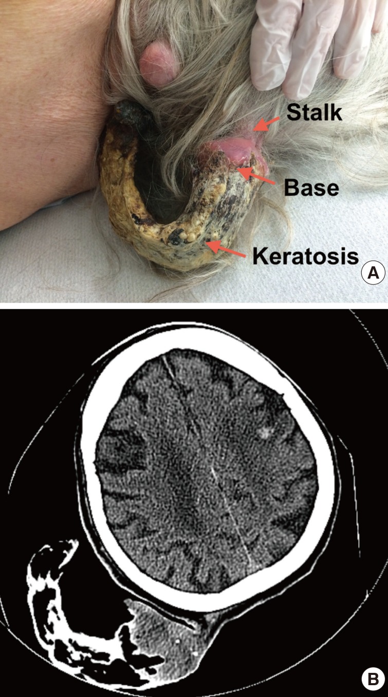
(A) Pedunculated giant cutaneous horn based at the occipital scalp. (B) Coronal 2-dimensional computed tomography image showing the lobular giant cutaneous horns base with calcifications attached to subcutaneous tissue by a 1.2-cm stalk.
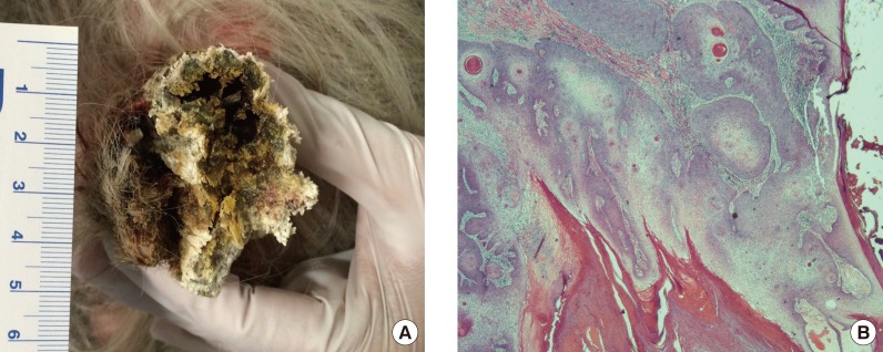
(A) Cross-section of the giant cutaneous horns (GCH) showing hollowing of conical component with surrounding parakeratosis, calcific debris and keratin. Ruler markings in centimeters. (B) H&E stained section (×100) of the GCH base with moderately differentiated squamous cell A B carcinoma.
Notes
We thank Dr. Janet Malowany, MD FRCP(C) in the Department of Laboratory Medicine at St. Joseph's Health Centre, Toronto, Ontario, Canada for reviewing the histopathology included in this case report.
No potential conflict of interest relevant to this article was reported.