Early Vascularized Fibular Grafts in Infants with Congenital Pseudarthrosis
Article information
Neurofibromatosis type I, also known as Von Recklinghausen disease, is one of the most common congenital disorders, with an incidence of 1 out of 3,000–4,000. It can be diagnosed on the basis of characteristic phenotypic manifestations, such as café-au-lait spots, axillary and inguinal freckling, neurofibromas, optic gliomas, Lisch nodules and distinctive osseous lesions. Osseous lesions occur in 2%–3% of neurofibromatosis type I cases, and are known to be caused by a lack of osteoblast function and the consolidation of osteoclast activity [1]. The most commonly affected region is the diaphysis of the tibia. Due to segmental disturbances in periosteal bone formation, anterior bowing occurs, and pathologic fractures eventually develop. In the first 2 years of life, pseudarthrosis may occur, which makes bone healing difficult due to persistent instability and progressive deformity in the bone. In patients with congenital pseudarthrosis, serious consequences such as amputation of the lower limb may be necessary if the condition is not adequately managed [2]. In this report, the authors describe the case of a vascularized fibular bone graft in a congenital pseudarthrosis patient.
A 15-month-old male patient fell from the sofa and did not stop crying. At the Department of Orthopedics of our institution, pseudarthrosis was diagnosed at the midshaft of the right tibia and fibula. Anterolateral bowing of the tibia was also seen. Café-au-lait spots were found on the posterior neck and both lower legs. Neurofibromatosis type I was diagnosed on the basis of genetic testing. Closed reduction and the application of an external fixation were performed at the Department of Orthopedics, and after 4 months, the external fixator was removed and bone union was identified. However, 2 days later, the same leg was fractured at the tibia (Fig. 1). The patient was referred to our department for a vascularized bone graft. The thick fibrous tissue around the pseudarthrosis of the tibia was incised and the tibia was exposed (Fig. 2) A fibula free flap 7 cm in length was planned to be placed at the diseased tibia. Using a saw, the tibia was partially resected to create a platform for the fibula bone graft 3.5 cm superior and inferior to the pseudarthrosis site. A 7-cm contralateral fibula free flap was harvested with a 5-cm pedicle. A skin paddle was elliptically designed based on 2 perforators originating from the peroneal artery (Fig. 3). The fibular bone graft was inserted into both slots of the tibia and internally fixated with plates and screws. End-to-side anastomosis was performed between the posterior tibial artery and the peroneal artery. Two concomitant veins were anastomosed in an end-to-end fashion. Endosteal bleeding of the fibular graft and capillary refill of the skin paddle was verified after anastomosis. The wound was closed in layers and a long leg splint was applied. Three months later, no abnormal changes were noted in X-ray imaging and no clinical wounds were observed (Figs. 4, 5).
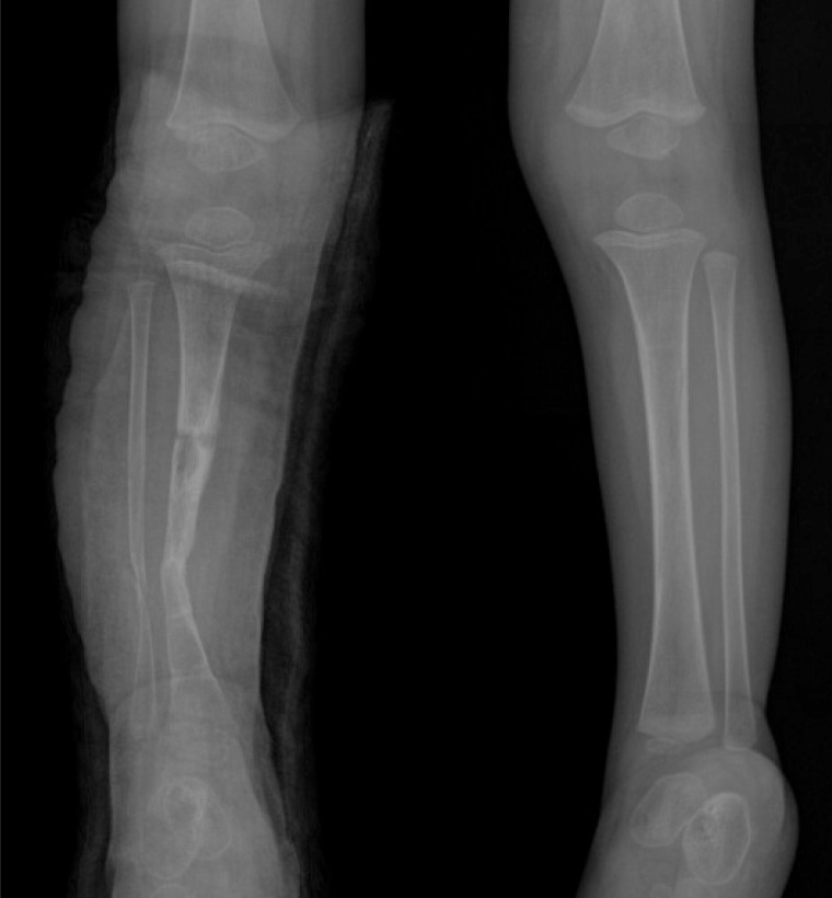
Preoperative radiographs of the patient. These preoperative radiographs of a 15-month-old male patient show pseudarthrosis of the right tibia and fibula, as well as a midshaft fracture of the tibia.
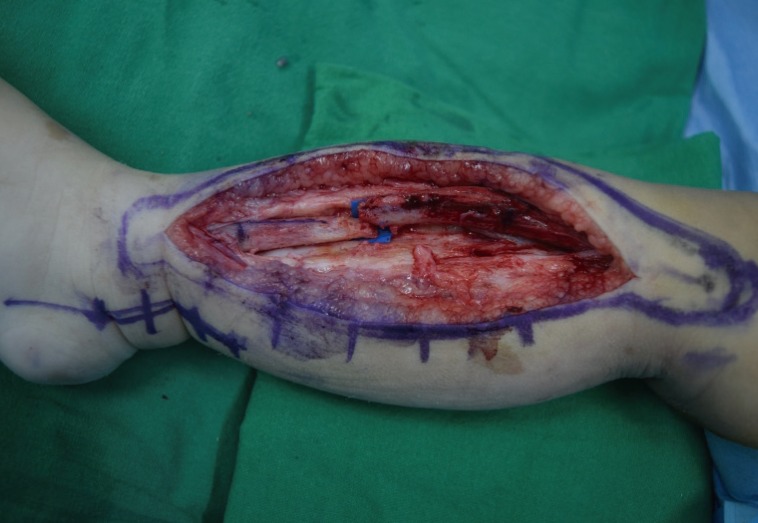
Intraoperative view. Pseudarthrosis and fracture of the right tibia midshaft can be seen. Anterolateral bowing of the tibia was also seen.
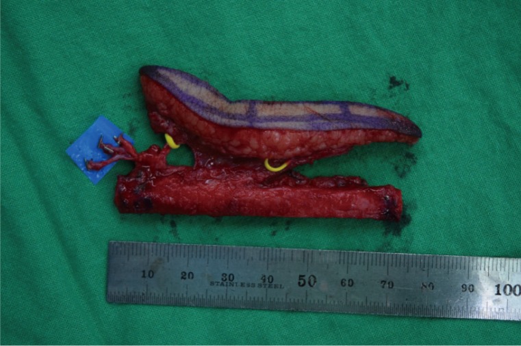
The free flap. A 7-cm contralateral vascularized osteocutaneous fibula free flap was harvested.
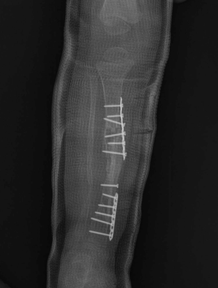
Three-month postoperative X-ray. The fibular bone graft was well inserted into both slots of the tibia and was internally fixated with plates and screws.
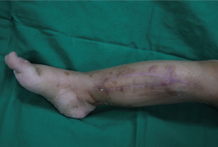
Results of the reconstruction 3 months postoperatively.
Conservative treatment is the most common strategy in patients with congenital pseudarthrosis. However, surgical intervention is required in case of non-union and recurrent fractures. The surgical therapy consists of resection of the diseased segment and bone grafting, followed by internal or external fixation [3]. In this case, the authors performed a vascularized bone graft with internal fixation. The diseased segment was minimally resected in order to act as a bone graft next to the vascularized fibula graft. Early intervention is advocated because delayed interventions increase the chance of a length discrepancy between the legs [4]. In young patients, sufficient consultation with the parents is necessary to obtain fully informed consent before proceeding with surgery.
Notes
No potential conflict of interest relevant to this article was reported.