The Role of Living Models in the Spending Review Era: How Do You Make the Most of a Rat?
Article information
Dear Sir,
I read with great interest the article “Comprehensive Analysis of Chicken Vessels as Microvascular Anastomosis Training Model” by Kang et al. [1].
In this paper, the authors discussed their evaluation of the anatomic characteristics of various chicken vessels as a training model.
The authors concluded that non-living chickens can provide various vessels with different anatomic characteristics, which can allow trainees the choice of an appropriate microvascular anastomosis training model depending on their purpose and skill level.
The increase in restrictions on animal use and the financial constraints of surgical training in recent years have led to the development and spread of many non-living animal models for microsurgery simulation. Such models are numerous and include rat cadavers, cryopreserved rat aortas, chicken and turkey wings, leaves and grape skin, human cadaver vessels, and different styles of plastic simulation materials [2,3].
Unfortunately, the abovementioned models do not allow the simulation of many factors that normally occur during microsurgical dissection and anastomosis, such as bleeding and vessel spasm [3,4]. Further, they prevent the surgeon from checking the anastomosis using a patency test. Therefore, living rat animal models are still indispensable.
In the existing literature, many living rat animal models have been reported, including some used for evaluating the performance of several exercises, such as flaps or transplants. However, all these models imply the need of a considerable number of animals since it is recommended to perform only 1 exercise on each vessel.
We have developed a simple method to make the most of a living model by performing several anastomoses in a row [5]. This allows researchers to exploit the same vessel to the maximum possible extent and leads to an amplification of the trainee surgeon’s mistakes, helping the novice surgeon to improve his/her surgical skills (Fig. 1).
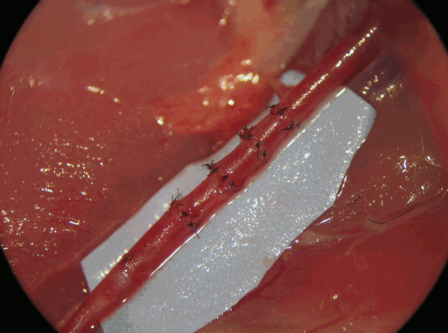
Series of anastomoses in a row conducted on a single femoral artery in a rat. The trainee performs several anastomoses in a row on both the femoral arteries, thereby using as few living animals as possible and minimizing animal wastage.
The rat model, particularly the rat femoral artery, is the most frequently used model in microsurgical training because of its ease of dissection, the optimal exposure of the vessel, and its dimensions.
This model is essential for learning to tackle the entire range of possible difficult situations in surgical practice.
Various factors contribute to the difficulty of performing several anastomoses in series: (1) incorrect stitch distribution along the margins, instead of an arrangement parallel to the blood flow direction; (2) twisting of a vessel between the anastomoses; (3) repeated traumatic pinching of the margins of a vessel; (4) increased platelet deposits downstream of each anastomosis; (5) limitations of the clamping space; (6) awkward surgical field; and (7) gradually increasing stress to the surgeon.
All these factors correspond to typical mistakes made by a novice surgeon, who does not realize, at the beginning of his/her training, the importance of avoiding them during a microsurgical suture. Further, this exercise incorporates error amplification, thereby enabling the surgeon to correct himself/herself.
The training of a novice microsurgeon is a step-by-step process. Starting from simple exercises such as end-to-end suturing of the femoral artery, the trainee should become able to perform more advanced procedures, such as rat kidney autotransplantation or the epigastric free flap. All the living models for microsurgical training reported thus far in the literature improve microsurgical skills by increasing the difficulty in vessel dissection or decreasing the vessel size, but none of them highlight the typical mistakes that a novice surgeon makes at the beginning of his/her training [2].
In our opinion, this model helps a novice surgeon to improve his/her skills with respect to anastomosis through an exercise involving the creation of 4 anastomoses in a row. This leads to an amplification of the trainee surgeon’s mistakes, which add up as the anastomoses are performed.
Performing serial anastomoses on the same artery avoids animal wastage by enabling the surgeon to work on the same vessel to the maximum possible extent.
In conclusion, the non-living model previously proposed by the authors [1] is a good method to prepare a trainee for a living model. However, the next step, in our opinion, should be utilizing a rat to perform several anastomoses in a row on both the femoral arteries, thereby using as few living animals as possible and minimizing animal wastage.
Notes
No potential conflict of interest relevant to this article was reported.