Interstitial Granulomatous Dermatitis with Granuloma Annulare-Like Pattern Following Liposuction
Article information
We hereby report a case of a 41-year-old woman with a history of liposuction of the right hip 6 months previously who presented to her dermatologist with bilateral recurring erythematous skin nodules. On palpation, 1 cm tender nodules were identified in the bilateral hip region in close approximation to the liposuction scars and a shave biopsy was performed from the lesions on the right side. The clinical differential diagnosis included panniculitis, tumid lupus erythematosus, and infection. A biopsy was performed and the histopathologic examination revealed an interstitial palisading granulomatous dermatitis with dermal mucin deposition confirmed by colloidal iron stain (Figs. 1–3). We examined multiple H&E sections, and there was no evidence of polarizable or non-polarizable foreign material. Special stains (Gomori Methenamine silver stain [GMS], Periodic acid-Schiff [PAS], and Acid-fast bacilli stain [AFB]) were negative for fungus and mycobacteria and culture studies performed also were negative.
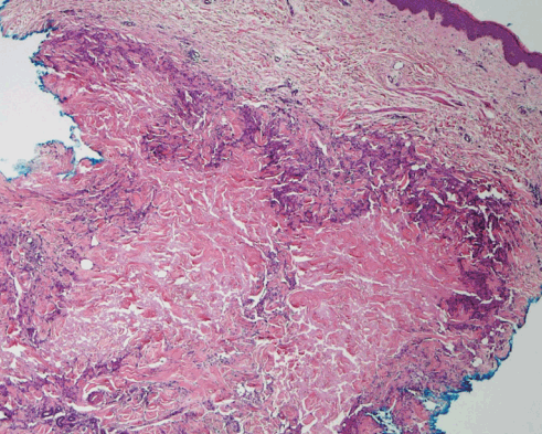
Interstitial granulomas (H&E, ×40). Section of skin showing dermal palisading granulomatous inflammation with central area of interstitial mucin deposition.
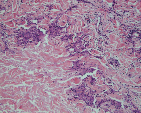
Interstitial granuloma with mucin (H&E, ×100). Sections demonstrating presence of palisading granulomatous dermatitis surrounding interstitial mucin deposits.
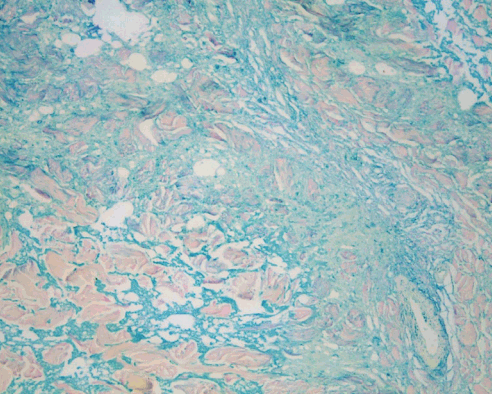
Special stain highlighting mucin. (Colloidal iron stain, ×100). Colloidal iron stain highlights the mucin (blue) in the central part of dermal interstitial granulomas.
In light of the patient’s history of liposuction immediately adjacent to the area of the nodules, we concluded that granulomatous dermatitis was induced by the liposuction procedure. There was no known history of any injectable material used in our case. The lesions have not recurred so far to the best of our knowledge.
Well-known adverse effects of liposuction include allergic contact dermatitis, seroma, post-inflammatory changes, and infection [1,2]. Rarely, a granuloma annulare-like reaction pattern secondary to foreign material has been reported after liposuction [3]. This is the first report to our knowledge of interstitial granulomatous dermatitis with a granuloma annulare-like pattern following liposuction, unassociated with any history of injected foreign material or foreign material by histologic examination.
Notes
No potential conflict of interest relevant to this article was reported.