Mechanical irritation by protruding bone: A possible cause of breast implant rupture
Article information
Abstract
Although breast implants have been in clinical use for almost 6 decades and have undergone considerable development during this time, implant rupture is still a dreaded long-term complication. Some obvious external factors, such as trauma, can lead to implant rupture, but many studies have reported a high rate of “spontaneous” implant rupture. Herein, we present two cases with the aim of raising awareness of a new possible cause of “spontaneous” implant rupture: mechanical irritation by bony protrusions.
INTRODUCTION
Although breast implants have been in clinical use for almost 6 decades and have undergone considerable development during this time, implant rupture is still a dreaded long-term complication. Patients with implant rupture usually present without any symptoms, which is referred to as silent implant rupture [1]. The symptoms and signs of rupture include obvious changes in breast shape, such as palpable insertions and pain. Symptomatic Implant rupture with the need to exchange the prosthesis occurs in 2% of patients who undergo breast augmentation [2,3]. After a physical examination, imaging including ultrasonography, magnetic resonance imaging (MRI), mammography, or computed tomography should be performed to verify the diagnosis. However, the rupture of an implant does not necessarily require the prosthesis to be exchanged. Silent ruptures can remain untreated with regular follow-up. Surgical intervention is only needed when the rupture causes symptoms [4].
In most cases of silicone implant rupture, direct or indirect trauma is reported [5]. Studies have shown that aged implants are predisposed to rupture, with a steadily increasing risk from 6 to 8 years after implantation [3,6,7]. Nevertheless, many studies have reported a high rate of “spontaneous” implant rupture. We suggest that these “spontaneous” ruptures could partially be triggered by endogenous factors, as described below.
CASES
Case 1
A 44-year-old female patient underwent total mastectomy of the right breast after being diagnosed with invasive ductal carcinoma. The sentinel biopsy was positive. Thus, axillary dissection of the lymph nodes was performed. She thereafter received chemotherapy, as well as radiation and hormone therapy.
The patient presented at our department after radiotherapy, chemotherapy, and hormone therapy with the desire for breast reconstruction. She appeared to be very athletic, without much subcutaneous fat tissue. Therefore, adequate autologous breast reconstruction was not a viable option. Despite the previous radiation therapy, the patient showed sufficient skin quality, so we decided to implant an expander. This was performed 1 year after ablation. Three months later, we exchanged it with an anatomic textured 275-mL implant with a moderate profile (Mentor Siltex, Mentor Worldwide LLC, Irvine, CA, USA).
Six months later, the patient presented again, showing signs of Baker grade IV capsular contracture in her left breast. There were no clinical signs of deflation of the implant. The patient did not recall any trauma to the chest or breast, either before the surgical procedures or after. On the patient’s request, ultrasound imaging was performed instead of MRI, and no implant rupture was visible.
The decision to perform a capsulectomy with implant exchange was made based on the clinical symptoms, with the patient’s consent. During the operation, the leakage of the implant became obvious (Fig. 1). Interestingly the area of the leakage was in direct contact with a protruding bony spur of the 6th rib. The implant was examined closely, and we noted that a small amount of silicone gel had leaked out through a small hole in the surface that had been in direct contact with the exostosis (Fig. 2). The diameter of the leakage was about 2 mm. After the bony spur was ablated using a bone rongeur, a capsulectomy was performed and a new implant was inserted. Over 5 years of follow-up, no new complications occurred.
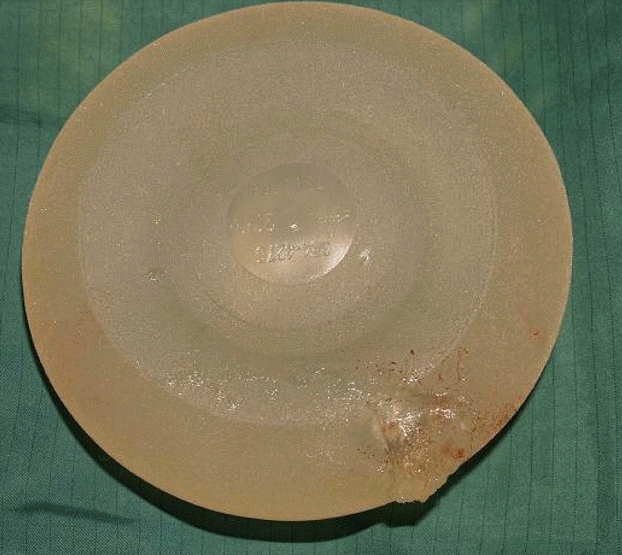
Implant leakage in case 1
Intraoperative picture of the explanted implant in case 1. Leakage in the lower right border of the implant is clearly visible.
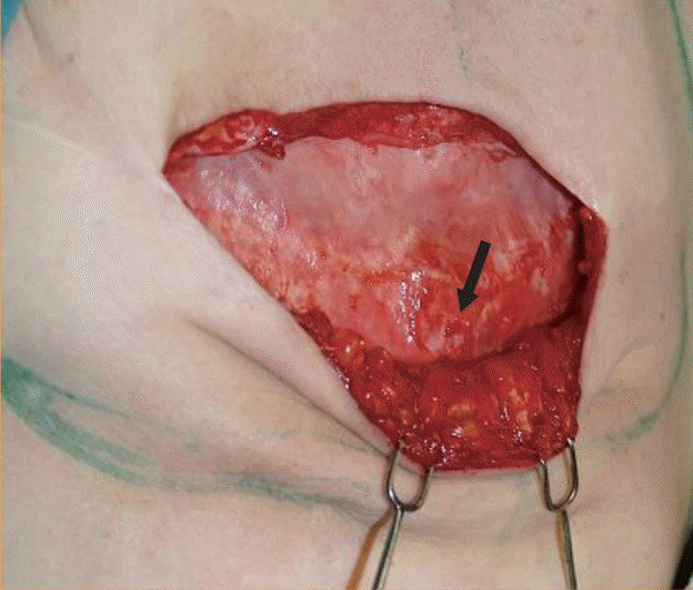
Intraoperative picture of the implant hole
The black arrow shows a protruding bony spur of the 6th rib, which had been in direct contact with the implant and may have caused the leakage.
Case 2
A 37-year-old female patient had undergone cosmetic augmentation. Five years after breast augmentation via a transaxillary approach, performed at another clinic, she presented at our department with pain in her left breast. She recalled minimal trauma. MRI was performed, and we verified implant rupture in the left breast. When explanting the ruptured implant (Mentor, Siltex, 300 mL, round, high profile) we found a bony spur of the 6th rib (Fig. 3). The bony spur was smoothened using a bone rongeur and the implant was replaced. During 3 years of followup, no further complications were noted.
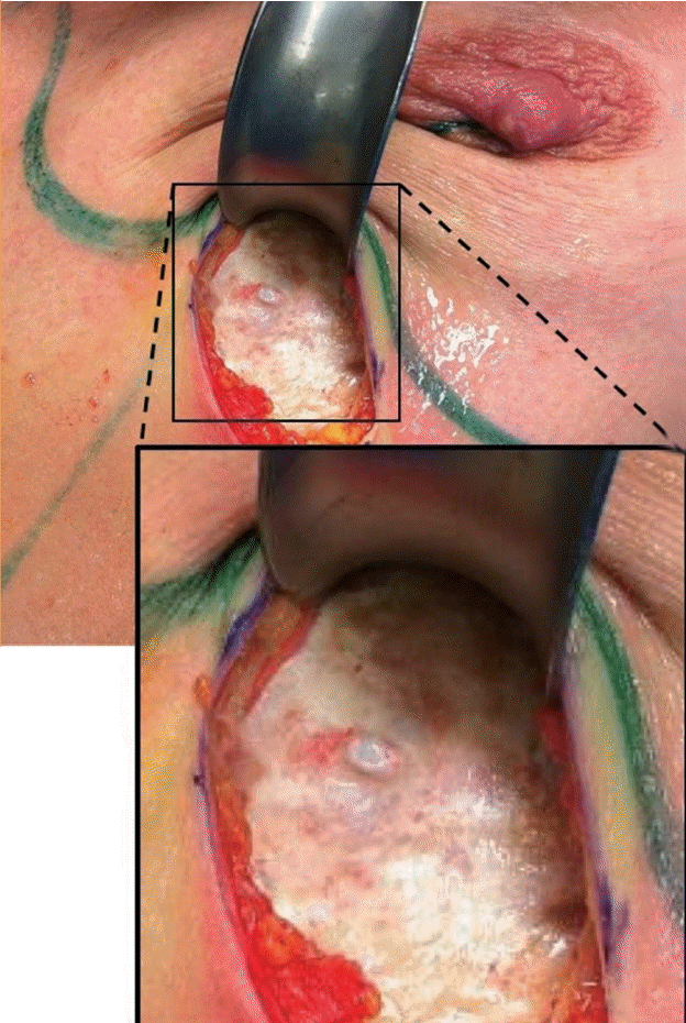
Intraoperative picture of case 2
Again, the exostosis was located on the 6th rib, and had been in direct contact with the implant, predisposing the implant to rupture.
DISCUSSION
In past decades, research has focused on the development of better implants. Nevertheless, surgeons also need to acknowledge individual anatomical characteristics, such as prominent ribs, in order to achieve optimal and safe long-term results. Various causes for the rupture or deflation of breast implants have been postulated in the literature. Although common causes, such as damage by surgical instruments, delamination, trauma, or manufacturing defects, have been described broadly in the literature [1,5,8-10], costal exostosis protruding from the outer part of the ribs into the subpectoral pocket is a potentially underrated threat to implant longevity. Difficult to recognize during the implantation of a prosthesis, prominent bone structures or exostoses of the ribs have the potential to lead to long-term complications requiring reoperation. As described in previous studies, long-term friction between the implant surface and the surrounding highly vascularized capsule can cause severe problems [11,12]. In case 1, we suspect that long-term friction was a predisposing factor for implant rupture.
Not only can pre-existing prominent bone structures cause implant-related problems, but prostheses have also been shown to enhance bone growth via mechanical stimulation. By enhancing the expression of osteogenic growth factors such as RUNX-2, PPAR-γ, and type I collagen in bone marrow mesenchymal stromal cells, mechanical irritation can lead to the formation of exostoses [13]. The growth of prominent structures can lead to enhanced, targeted pressure on the implant, and in combination with additional external forces, this may cause implant rupture. We suspect that implant rupture in case 2 was caused by this mechanism. Local bony hypertrophy led to enhanced pressure at a single point. Combined with slight trauma, this resulted in rupture of the implant. Therefore, it is crucial to visualize the entire implant pocket when exchanging a ruptured implant. When an exostosis is found, a smoothing ostectomy is necessary to reduce the risk of mechanical implant rupture.
As presented in two cases, direct contact of the implant with an underlying bone has the potential to trigger local bone formation. The arising exostoses have the potential to enhance punctual pressure on the implant, thereby triggering a rupture. Therefore, when changing an implant, attention should always be paid to the surface of the chest wall and the surroundings, as well as to the implant itself. The occurrence of punctual ruptures can indicate damage during implantation, but may also be a sign of local bone hypertrophy. Therefore, visualization of the entire implant pocket and the ablation of hypertrophic bony structures are essential to eliminate the risk of mechanical irritation in the long-term.
Notes
No potential conflict of interest relevant to this article was reported.
Patient consent
The patients provided written informed consent for the publication and the use of their images.