Regraft from Amputated Forehead Tissue of a 4×7 cm Skin Defect Containing Bony Defects
Article information
It is well established that the diameter of skin defects should not exceed 1 cm for the survival of the composite tissue graft [1,2], and that central necrosis is unavoidable if the diameter of the defects is greater than 2 cm. In addition, it is recommended that the composite tissue graft be performed no later than within four hours after the onset of injury [1,2]. This is also accompanied by the use of such methods as a pressure dressing with a tie-over for successful engraftment. Cryotherapy is also used to lower the metabolic rate [3]. It has also been reported that vascular anastomosis can be an effective treatment modality if the size of a skin defect exceeds 2 cm [1,2].
We treated a 4×7 cm skin defect on the frontal region containing bony defects of approximately 2×3 cm using a regraft from amputated soft tissue of the forehead. Thus, we obtained a good treatment outcome without using previously reported treatment methods such as a tie-over dressing although vascular anastomosis could not be performed in our case. Here, we report on our treatment outcomes.
In July 2009, a 52-year-old woman visited an emergency room with a chief complaint of a soft tissue defect of 4×7 cm containing bony defects approximately 2×3 cm in size extending over the forehead and scalp because the patient sustained an injury from a deadly metal weapon by her husband (Fig. 1A). The injury resulted in the amputation of soft tissue of the forehead and scalp. At the time of the emergency room visit, the patient also presented with a small bowel perforation due to a stab injury in the abdomen. After undergoing an emergency laparotomy in the Department of Surgery for approximately 3 hours and receiving treatment at an intensive care unit, the patient was referred to us for further evaluation and treatment.
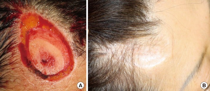
(A) At the time of onset of injury, the patient had a skin defect of 4×7 cm containing a bony defect of 2×3 cm. In the center of the bony defect, the medulla was exposed. (B) Approximately 21 months postoperatively, during an outpatient visit, the patient showed results of such a successful engraftment of the graft tissue that some hairs even grew at the site of transplantation. In addition, the skin color at the site of transplantation showed no notable differences from the adjacent tissue. Furthermore, the patient had no notable complications.
At the time of referral to us, the patient had not presented with the amputated tissue. Therefore, following conservative treatment, it was decided that a skin graft or flap surgery would be performed once granulation tissue had formed. During the monitoring of the clinical course, the patient's caregiver found the amputated soft tissue of the forehead region, approximately 7 hours after the injury at the location where the patient sustained the injury (Fig. 2).
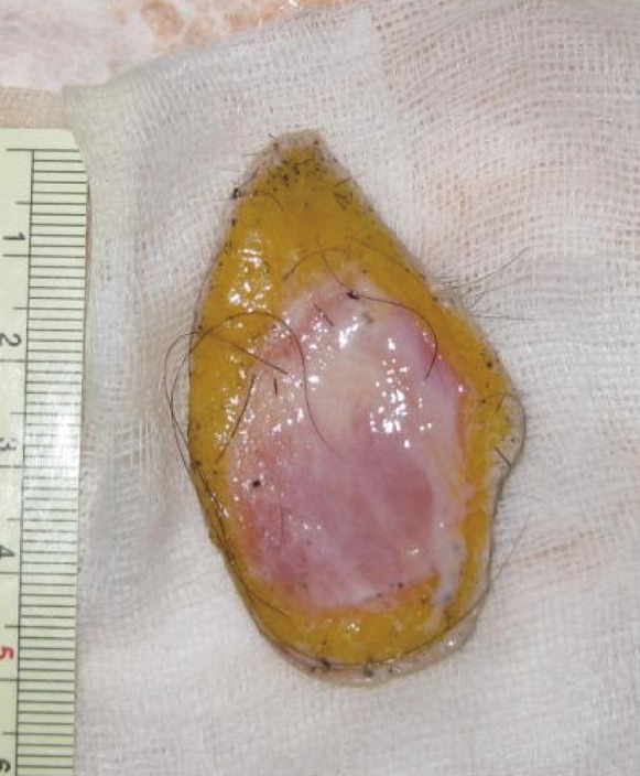
Amputated forehead and scalp soft tissue. The amputated tissue was severely contaminated.
We had attempted to perform a microvascular anastomosis, but we could not because the anesthesiologist judged that the patient could not undergo general anesthesia again.
However, if treatment were further delayed, the patient would have been at risk of increased damage. Therefore, we performed the regraft of the amputated tissue under local anesthesia 2 hours later.
The size of the amputated tissue was huge, but vascular anastomosis was impossible; therefore, we decided to remove the bone chips from the amputated tissue and perform massive defatting to reduce the metabolic rate.
Efforts were made to accurately fit the dermal layer between the skin defects as accurately as possible using Prolene 6-0 sutures. Immediately after transplantation, we did not perform a tie-over or compression dressing. However, we performed simple dressing changes 3 times a day to closely monitor the status of the flap, and thereby ensured that blood clots did not form on the incision line to prevent hematoma.
From postoperative day 6 on, a pink color began to gradually appear at the transplantation site. This was followed by suture removal without any notable aggravation to the patient's condition (Figs. 3, 4).
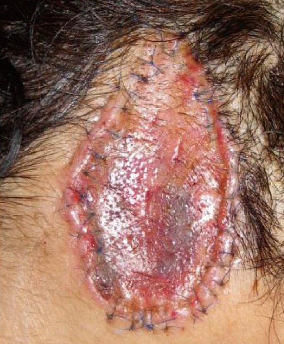
On postoperative day 10, the recipient tissue gradually began to take on a pink color
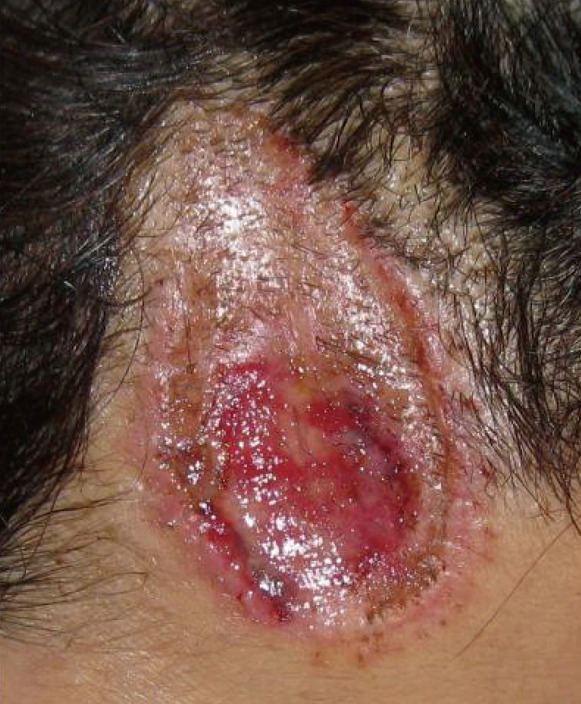
On postoperative day 12, the stitches were removed.
In general, a successful engraftment cannot be predicted if the diameter of a skin defect is greater than 1 cm. It is known that a successful engraftment cannot be achieved if at least 4 hours has elapsed from the time of injury without any specific preservation processes such as freezing the amputated tissue following the onset of injury [1,2]. Furthermore, there are recommended methods for increasing the likelihood of engraftment success such as a compression dressing with a tie-over dressing and cryotherapy that lowers the metabolic rate [3].
In our case, the skin defect had a size of approximately 4×7 cm, and this was accompanied by the presence of bony defects with a size of approximately 2×3 cm. Besides, approximately 7 hours had elapsed from the onset of injury until the amputated soft tissue of the forehead region was found, and the tissue was severely contaminated.
Additionally, the patient had a poor systemic status following an emergency laparotomy in the Department of Surgery for the management of small bowel perforation, and was receiving treatment in the intensive care unit. Vascular anastomosis could not be performed for surgical management due to the patient's condition. Therefore, we attempted to accurately fit the dermal layer between the skin defects, and thus attempted to induce angiogenesis as promptly as possible within the dermal plexus.
Approximately 21 months postoperatively, during an outpatient visit, the patient showed results of such a successful engraftment of the graft tissue that some hairs even grew at the site of transplantation. In addition, the skin color at the site of transplantation showed no notable difference from the adjacent tissue. Furthermore, the patient presented with no other complications than depression at the site of injury (Fig. 1B).
This case shows that even though the skin defect was greater than 1 cm, a substantial period of time elapsed prior to the surgery, and the period of ischemic condition was prolonged, we were able to perform a regraft from the amputated tissue instead of vessel anastomosis. We obtained such a successful treatment outcome that entire engraftment of the amputated tissue as achieved without partial necrosis.
Notes
No potential conflict of interest relevant to this article was reported.