Epithelial-mesenchymal transition in keloid tissue
Article information
Fibroblasts at the wound site are recognized as the primary drivers of scar formation. They differentiate into myofibroblasts, the key mediators of fibrosis, which are responsible for collagen deposition and wound contraction. Repair processes cease when epithelialization is completed in normal wounds, whereas in keloid wounds, they may continue and result in excessive accumulation of unorganized extracellular matrix, forming problematic scars.
In addition to resident mesenchymal cells, fibroblasts and myofibroblasts are thought to be derived from multiple sources, including epithelial-mesenchymal transition (EMT) [1]. During this process, epithelial cells experience intercellular and intracellular changes, including dissociation of junctional complexes, loss of apical-basolateral polarity, and repression of epithelial markers. As a result, epithelial cells lose many of their properties and take on characteristics of mesenchymal cells.
To investigate this process, an immunofluorescence assay using both epithelial and mesenchymal markers was performed using keloid tissues obtained from the anterior chest wall of a 70-year-old male patient (Fig. 1). After fixation, cells were incubated with primary antibodies against E-cadherin and vimentin (Abcam, Cambridge, UK), and subsequently labelled with fluorescently-tagged secondary antibodies. Counterstaining with 4’,6-diamidino-2-phenylindole (DAPI) (VECTOR Laboratories, Burlingame, CA, USA) was also performed.

Patient with a keloid. A 70-year-old male with a keloid on the post-sternotomy area suffered from itchiness, pain, and skin tightening. The keloid was surgically excised intralesionally.
Cells were visualized using an LSM 700 Carl Zeiss confocal microscope (Carl Zeiss MicroImaging, Thornwood, NY, USA). E-cadherin and vimentin expression were simultaneously observed at the dermo-epidermal junction in keloid tissue, indicating the occurrence of EMT (Fig. 2).
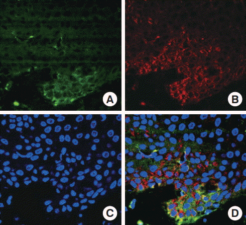
Epithelial-mesenchymal transition (EMT) characteristics in keloid tissue. (A) Expression of the epithelial marker E-cadherin (green) and (B) the mesenchymal marker vimentin (red) was observed via immunofluorescence. (C) DAPI counterstain of nuclei (blue). (D) Co-expression of E-cadherin and vimentin at the dermo-epidermal junction was observed, indicating the occurrence of the EMT process (A-D, magnification ×630).
Although further investigations with additional keloid specimens are needed, we hope that our study yields some insight into the mechanisms of keloid formation.
Notes
No potential conflict of interest relevant to this article was reported.
Ethical approval
The study was approved by the Institutional Review Board of Yonsei University Medical Center (IRB No. 4-2017-0259) and performed in accordance with the principles of the Declaration of Helsinki. Written informed consent was obtained.
Patient consent
The patient provided written informed consent for the publication and the use of his image.