Synchronous reconstruction of both the upper and lower eyelids with a temporoparietal fascial flap
Article information
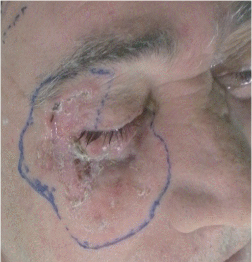
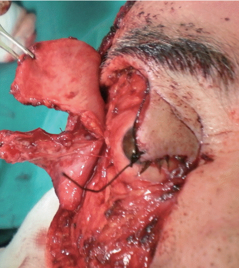
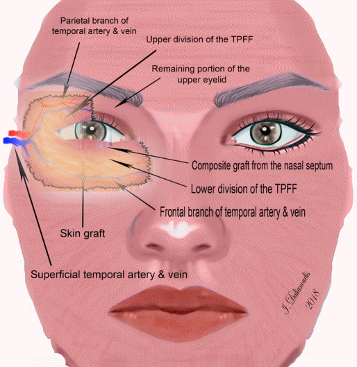
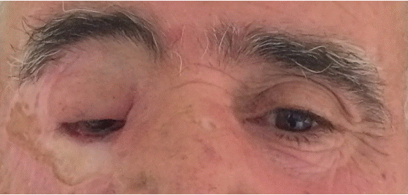
The simultaneous reconstruction of upper and lower eyelid defects, while preserving the seeing eye, presents a difficult task. Most cases involving such defects occur after burn injuries and tumor resections [1]. We present the case of a 55-year-old man, who provided consent and authorization for this report, with neglected basal cell carcinoma of both the upper and lower eyelids in the right eye and 10/10 visual acuity (Fig. 1). After oncological resection, defects were encountered in the total lower eyelid and half of the upper eyelid. A temporoparietal fascial flap (TPFF) was used to reconstruct both the upper and lower eyelids after an incision was made between the frontal and parietal branches of the temporal artery (Fig. 2). A composite graft from the nasal septum was sutured to the lower division of the TPFF and was used to reconstruct the posterior lamella, while the anterior lamella was reconstructed with a full-thickness skin graft (Fig. 3). During the 1-year follow-up period, no complications were encountered, except reduced upper eyelid motion, and the patient’s visual acuity was similar before and after reconstructive surgery (Fig. 4). The patient was satisfied with the postoperative aesthetic results and refused any further refinements. Even though the TPFF is a well-known flap, as far as we know, such reconstructions are generally multistage [2]. To our knowledge, only one report has described functional reconstruction of both eyelids, the eyebrow, and the lacrimal drainage system in a single-stage procedure by pre-dividing a TPFF into three divisions [3]. The described method offers an efficient, effective alternative for eyelid reconstruction in a single-stage procedure.

Planned oncological resection. A 55-year-old man with basal cell carcinoma of both the upper and lower eyelids that had been neglected for 8 years, with a functional eye and 10/10 visual acuity at presentation. The oncological resection involved the entire lower eyelid and half of the upper eyelid.

The temporoparietal fascial flap (TPFF). The TPFF after elevation and division into an upper and lower part following the course of the frontal and parietal branches. The two parts were used for the reconstruction of the upper and lower eyelids, respectively.

Schematic representation of the surgical procedure. In the sketch, we can see the setting of the temporoparietal fascial flap (TPFF) and the basic components of the procedure. The placement of the composite graft (cartilage+mucous) is highlighted. The course of the superficial temporal artery with its respective branches and the positioning of the two parts of the TPFF are simulated. Finally, the TPFF was covered with a full-thickness skin graft.

Presentation of the case 1 year after reconstruction. Postoperative results after 1 year of follow-up. The patient was satisfied with the postoperative aesthetic results. He experienced reduced upper eyelid motion that partially obscured his vision, but refused any further refinements.
Notes
No potential conflict of interest relevant to this article was reported.
Ethical approval
The study was performed in accordance with the principles of the Declaration of Helsinki. Written informed consent was obtained.
Patient consent
The patient provided written informed consent for the publication and the use of his images.
