Squamous cell carcinoma identified in a thick-walled epidermal cyst with a recurrent ulcer
Article information

Epidermal cysts are common skin tumors. In order to achieve cost-effectiveness, routine biopsy is not recommended in the setting of plastic surgery [1]. We report a case of squamous cell carcinoma (SCC) confirmed in a residual wound after resection of a cutaneous cyst. A 46-year-old male patient underwent local resection of a cystic lesion in the left nasal alar region 3 weeks before at a local medical center without biopsy. He developed tenderness, fever, and erythema over the wound, and visited our hospital (Fig. 1A). While the planned excision was performed, a 0.8×0.9-cm-thick white capsule was observed (Fig. 2). Therefore, a biopsy including skin tissue was performed. A SCC was then diagnosed (Fig. 3), and additional resection with a 5-mm safety margin was performed. During the 18-month follow-up period, no recurrence was observed (Fig. 1B). Owing to the rarity of SCC arising from epidermal cysts, the nature and mechanism of this phenomenon remains uncertain. Malignant change of an epidermal cyst is suspected when a chronic wound shows a sudden increase in size and ulceration. In such cases, other authors have suggested that biopsy and complete excision with a pathological examination should be performed [2]. According to a recent study, reported cases of SCC from epidermal cysts have increased [3]. Thus, to exclude malignant tumors, a biopsy is recommended when epidermal cysts with recurrent ulcers show wall thickening.
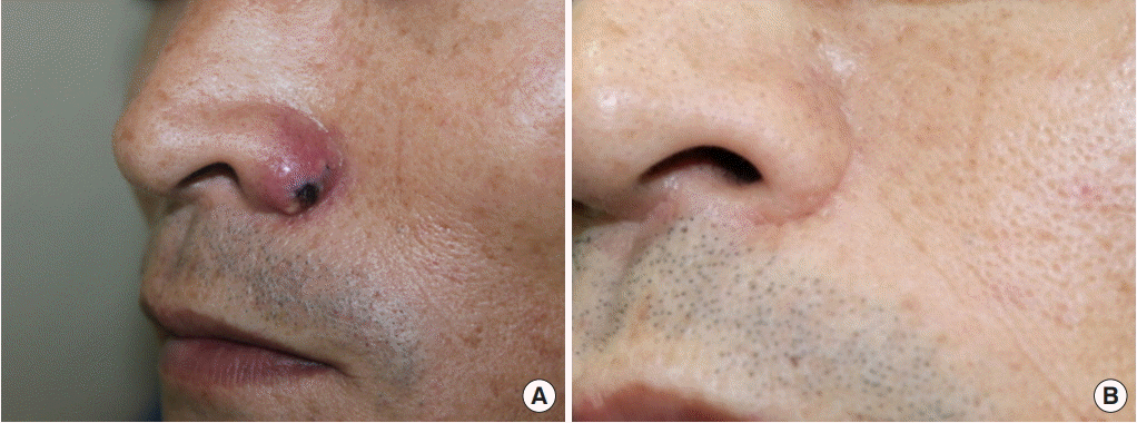
(A) The patient at his first visit to our hospital. He had developed tenderness, febrile sensation, and erythema on the wound in the left nasal alar region. (B) Six-month postoperative follow-up. The patient shows a well-healed state in the nasal alar region without recurrence.
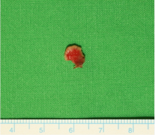
A biopsy specimen. A 0.8×0.9-cm-thick white capsule was observed.
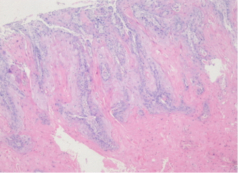
Cells showing mild to moderate atypia with abundant pink cytoplasm and carcinoma with well-developed keratinization, showing infiltration downward. These are typical findings of squamous cell carcinoma (hematoxylin and eosin stain, ×40).
Notes
No potential conflict of interest relevant to this article was reported.
Ethical approval
The study was approved by the Institutional Review Board of Yeungnam University Hospital (IRB No. YUMC 2018-09-026) and performed in accordance with the principles of the Declaration of Helsinki. Written informed consent was obtained.
Patient consent
The patient provided written informed consent for the publication and the use of his images.