Surgical excision of a giant soft fibroma of the labia majora covered with a pudendal artery perforator flap
Article information
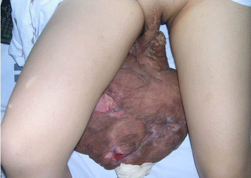
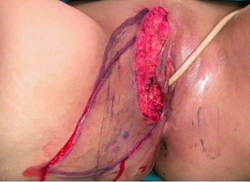
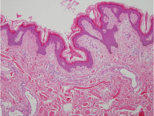
Tumors of the vulva are very rare, and only a few cases have been reported. Soft fibroma is the most common benign tumor of the genital area; it is a benign proliferation that has multiple forms and is usually less than 5 cm in size [1]. It frequently occurs on the torso or face, in locations such as the eyelids, neck, or axilla, and is rarely found on the vulva [2]. Initially, the tumor is often asymptomatic, but soft fibromas on the vulva can cause extreme social withdrawal, urinary symptoms, and bleeding due to ulceration after years of growth. Vulvar tumors are usually treated by surgical excision and, unless confirmed to be malignant, they typically do not present complications [3]. A 32-year-old nulliparous patient presented to the gynecology department as an outpatient with a painless, soft-to-firm, pedunculated mass on the labia majora. The tumor measured 60 cm in length, 50 cm in width, and 15 cm in thickness, and contained an ulcerative skin defect 4 cm in diameter. The mass had been progressively growing over the past 15 years (Fig. 1). A general and speculum examination revealed no other abnormalities. The patient underwent total excision of the mass from the stalk encircling the base of the attachment and a coverage operation with a musculocutaneous flap using internal pudendal artery perforators under general anesthesia (Fig. 2). A histopathological examination showed fibrous tissue without atypia, consistent with soft fibroma (Fig. 3). The patient was discharged 2 weeks after surgery without complications. During the follow-up period, no additional complications were encountered (Fig. 4).

Clinical photograph of a huge mass on the patient's labia majora. The tumor measured 60 cm in length, 50 cm in width, and 15 cm in thickness, with an ulcerative skin defect 4 cm in a diameter.

Intraoperative clinical photograph of the musculocutaneous flap. A musculocutaneous flap using internal pudendal artery perforators was designed after total excision of the giant soft fibroma.

Histopathological examination. Hyperplastic epidermidis with hyperkeratosis and acanthotic features consistent with soft fibroma (H&E, ×100).

Clinical photograph of the wound postoperatively. The patient was discharged without any complications, and no additional complications were encountered during 12 months of follow-up.
Notes
Conflict of interest
No potential conflict of interest relevant to this article was reported.
Ethical approval
The study was performed in accordance with the principles of the Declaration of Helsinki. Written informed consent was obtained.
Patient consent
The patient provided written informed consent for the publication and the use of her images.
Author contribution
Conceptualization, methodology: Jung SN. Methodology and visualization: Seo BF. Project administration: Choi JY. Data curation, writingoriginal draft: Cho JT. Writing-review & editing: Choi J, Choi JY.
