Reconstruction of Medial Orbital Wall Fractures without Subperiosteal Dissection: The “Push-Out” Technique
Article information

Abstract
Background
Various surgical methods for repairing medial orbital wall fractures have been introduced. The conventional technique requires total separation of the displaced orbital bones from the orbital soft tissues. However, subperiosteal dissection around the fracture can cause additional damage. The aim of the present study is to introduce a method of reconstructing medial orbital wall fractures without subperiosteal dissection named the “push-out” technique.
Methods
Six patients with post-traumatic enophthalmos resulting from an old medial orbital wall fracture and 10 patients with an acute medial orbital wall fracture were included. All were treated with the push-out technique. Postoperative computed tomography (CT) was performed to assess the correct positioning of the implants. The Hertel scale and a comparison between preoperative and postoperative orbital volume were used to assess the surgical results.
Results
Restoration of the normal orbital cavity shape was confirmed by examining the postoperative CT scans. In the old fracture group, the median orbital volume of the fractured side was 29.22 cm3 preoperatively, and significantly improved postoperatively to a value of 25.13 cm3. In the acute fracture group, the median orbital volume of the fractured side was 28.73 cm3 preoperatively, and significantly improved postoperatively to a value of 24.90 cm3. Differences on the Hertel scale also improved, from 2.13 mm preoperatively to 0.25 mm postoperatively in the old fracture group and from 1.67 mm preoperatively to 0.33 mm postoperatively in the acute fracture group.
Conclusions
The push-out technique can be considered a good alternative choice for old medial orbital wall fractures with posttraumatic enophthalmos, acute medial orbital wall fractures including large fractured bone segments, and single-hinged greenstick fractures.
INTRODUCTION
Orbital fractures are commonly seen with midfacial trauma. In the past, isolated blowout fractures of the medial orbital wall were often overlooked because they are less symptomatic than inferior orbital wall fractures. As computed tomography (CT) for facial trauma patients has become popular, the reported incidence of medial orbital wall fractures has increased in recent years.
Reconstruction of medial orbital wall fractures is a challenging problem for surgeons because of the narrow operative field with complicated anatomical structures. In a limited space, many structures conjoin to form the medial orbit. The medial orbital wall is formed by the lamina papyracea of the ethmoid, lacrimal, sphenoid, and frontal bones, along with the anterior and posterior ethmoidal arteries and the optic nerve. The goal of treatment in patients with a medial orbital wall fracture is appropriate reconstruction of the deformed orbital structure to restore the balance between the volume of the orbital soft tissues and the orbital cavity.
The most well-known surgical technique, onlay implantation, involves the onlayed coverage of bony defects with implant insertion onto the fracture site. The conjunctival approach is very helpful for performing this procedure. The medial transcaruncular approach leaves no detectable scars, leading to aesthetically pleasing results [1]. Many surgeon have performed this technique, but it is sometimes difficult for the surgeon to maintain an optimal view. The limitation of this approach is that the operative field is less visible, making it difficult to proceed with surgical procedures such as dissection and the insertion of large alloplastic implants into this complex fractured area [2]. This conventional onlay technique needs an area of subperiosteal dissection that is wider than the measured fracture area. It requires that the displaced bone segments be separated from the soft tissues [2]. To overcome those obstacles, the authors of the present study previously introduced the inlay implanting method [2], which involves inserting alloplastic implants layer by layer into the ethmoidal sinus of the fractured site. This inlay implanting technique needs minimal subperiosteal dissection and can be used in comminuted medial orbital fractures, allowing adequate reconstruction of orbital continuity without the fear of damaging the optic nerve.
However, both of onlay and inlay implantation require subperiosteal dissection, which may lead to serious complications, such as bleeding or a nerve injury. Particularly in patients with old fractures with posttraumatic enophthalmos, the scar tissue surrounding the fracture site could be adhered with vessels, nerves, or extraocular muscles. Subperiosteal dissection around the fracture site can therefore be dangerous. The adhesion is random and unpredictable in a narrow operative field, making subperiosteal dissection more difficult.
Bone repositioning without subperiosteal dissection can resolve this problem. This method, in which the fractured bone segment is repositioned into the original position, is considered ideal and physiologic in that it uses the fractured bone segment itself. The bone repositioning method was introduced previously, using endoscopy or a balloon catheter [3,4]. Recent advances in endoscopic techniques have allowed fractured medial orbital walls to be repositioned with excellent visualization and complete dissection of the circumference through the endoscopic transnasal approach. However, endoscopic equipment is required, and the surgeon must be quite skillful at endoscopy.
In light of the above considerations, we sought to find a simple and minimally invasive reconstruction method without subperiosteal dissection. Herein, we describe a modified inlay implanting method including medial orbitotomy, referred to as the transcaruncular “push-out” technique.
METHODS
Patients
Among the patients who were diagnosed with a unilateral medial orbital wall fracture between August 2013 and December 2014, 16 underwent surgery for correction of a medial orbital wall fracture. We began this study after all patients received information on the purpose and possible side effects of examinations and signed an informed consent. All authors have read the Declaration of Helsinki and have followed the guidelines in this study. Among the 16 patients, 6 suffered from posttraumatic enophthalmos with an old medial orbital wall fracture (the old fracture group), and 10 had an acute medial orbital wall fracture (the acute fracture group). The indications for the operation were as follows: reduced visual acuity, diplopia, abnormal extraocular muscle movement (EOM), severe fracture (size of fracture greater than 2 cm2 on CT scans) or enophthalmos with a difference of more than 2 mm between the injured and contralateral orbits.
Preoperative evaluation
Before the operation, all patients were evaluated for the position and symmetry of the orbits, vision, diplopia, extent of EOM, any accompanying ocular diseases, or any other symptoms. If necessary, patients were evaluated by an ophthalmologist who understood the operation. The presence of enophthalmos was assessed with a Hertel exophthalmometer (Oculus Inc., Wetzlar, Germany). Measurements were performed 3 times by a single surgeon in each patient, and the mean of the 3 values was used. The orbital volume was calculated using an Aquarius workstation (iNtuitionaquarius, ver. 4.4.6., TeraRecon, San Mateo, CA, USA). In order to plan a precise operation, we analyzed the axial and coronal views of CT scans to assess the status of orbital structures, such as the size of the bony defects and the degree of orbital tissue herniation. We decided to use our push-out method when there was no soft tissue entrapment.
Surgical technique
The preoperative design was marked between the caruncle and plica semilunaris conjunctivae for a transcaruncular incision. The marked area was incised, and through this incision, minimal subperiosteal dissection proceeded up to the bony rim of the medial orbit. Subsequently, a bony hole was created on the anterior wall of the medial orbital rim using a drill. Then, medial orbitotomy was performed with an osteotome and mallet. We inserted a thin periosteal elevator into the ethmoidal sinus via the medial orbitotomy approach and detected the displaced bone segment. When reaching for the fractured segment, we gently placed pressure on the periosteal elevator and gradually corrected the fracture (Fig. 1). The process we have just described is referred to as the “push-out” technique. We then inserted and placed porous polyethylene implants in small pieces into the ethmoidal sinus. Implants were placed under the fractured bone segment to counteract the tendency for displacement to recur. To confirm ocular mobility, the forced duction test with toothed forceps was repeatedly conducted. Finally, primary closure at the incision was performed and postoperative dressing with an ointment was performed.
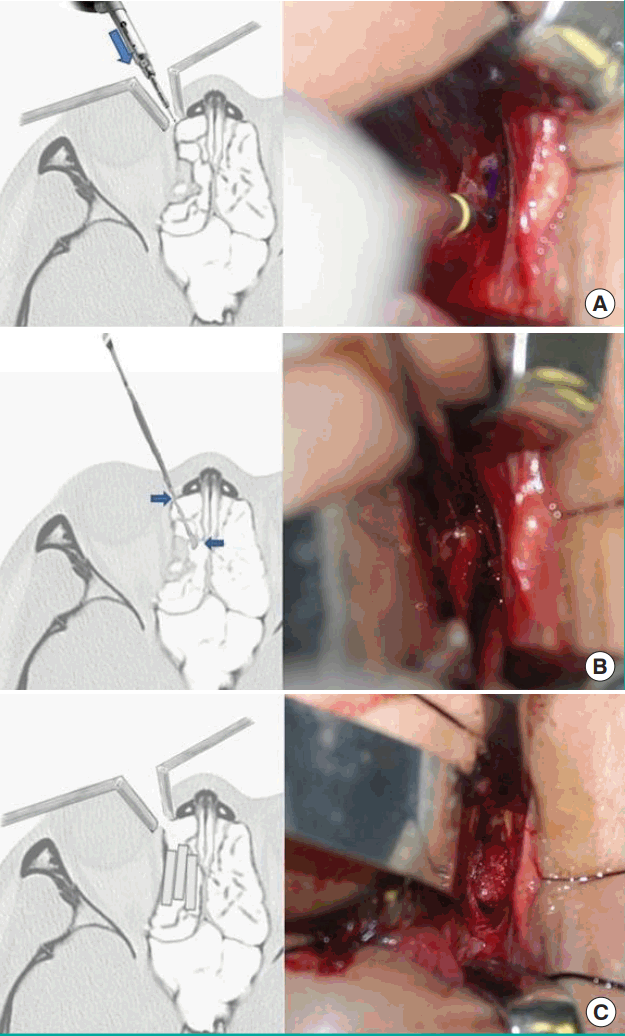
Intraoperative photograph of the “push-out” technique
(A-C) Schematic drawing and intraoperative photograph of the “push-out” technique for reconstruction of the medial orbital wall. (A) The blue arrow indicates the insertion of a thin periosteal elevator into the ethmoidal sinus via the medial orbitotomy approach. (B) The blue arrows indicate gentle and gradual correction of the fractured segment by the periosteal elevator.
Postoperative evaluation
We had a median follow-up period of 9 months, ranging from 6 to 15 months. A Hertel exophthalmometer (Oculus Inc.) was used to assess the extent of enophthalmos. A postreconstructive functional assessment was performed as well, with an evaluation of diplopia and EOM limitations. In order to assess the aesthetic outcomes, preoperative and postoperative CT scans were reviewed. Postoperative orbital volumetric measurements were made with an Aquarius workstation.
Statistical analysis
The Wilcoxon signed-rank test was used to analyze perioperative differences in the orbital volumes measured using the Aquarius workstation and the Hertel scale between the 2 orbits. P-values <0.05 were considered to indicate statistical significance. All statistical analyses were performed using SPSS ver. 20.0 (IBM Corp., Armonk, NY, USA).
RESULTS
All 16 medial orbital wall fracture patients in the current study were adequately treated. Restoration of the normal orbital cavity shape was identified on the axial and coronal views of postoperative CT scans in all cases. No complications occurred, except for 1 patient in whom some of the comminuted fracture was missed on preoperative imaging; this patient underwent a revisional operation because of newly developed diplopia after the operation. The patient who underwent the revisional operation showed improved diplopia symptoms. In patients with an old medial orbital wall fracture, the median (interquartile range, IQR) orbital volume of the fractured side was 29.22 cm3 (28.31–29.98 cm3) preoperatively, which significantly improved to a postoperative value of 25.13 cm3 (24.41–25.80 cm3). In patients with an acute medial orbital wall fracture, the median (IQR) orbital volume of the fractured side was 28.73 cm3 (27.86–29.49 cm3) preoperatively, which significantly improved to a postoperative value of 24.90 cm3 (24.33–25.66 cm3) (Table 1). In patients with an old medial orbital wall fracture, the median (IQR) difference on the Hertel scale was 2.13 mm (2.06–2.22 mm) preoperatively, which improved to 0.25 mm (0.22–0.27 mm) postoperatively. In patients with an acute medial orbital wall fracture, the difference on the Hertel scale was 1.67 mm (1.61–1.74 mm) preoperatively, improving to 0.33 mm (0.31–0.36 mm) (Table 2).
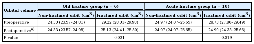
Orbital volume changes measured using the Aquarius workstation
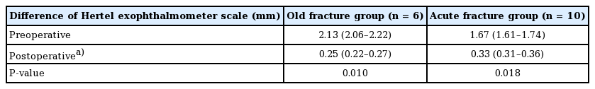
Changes on the Hertel scale measured by a Hertel exophthalmometer (Oculus Inc.)
Case descriptions
Case 1
A 38-year-old woman visited the outpatient clinic with enophthalmos on the left side. She had been in a car accident 1 year ago. At the time of the visit, she did not show any functional problems related to diplopia, impaired vision, or abnormal ocular movements. The medial orbital wall fracture and herniated orbital contents on the left side were identified on a CT scan. She underwent an operation using the push-out technique. The herniated orbital contents and the fractured bone segment were restored to the original anatomical position. Three porous polyethylene implants measuring approximately 15 mm×6 mm, with a thickness of 3 mm, were placed in the ethmoidal sinus layer by layer. Postoperative CT imaging revealed an adequately reconstructed medial orbital wall (Fig. 2). No complications were observed.
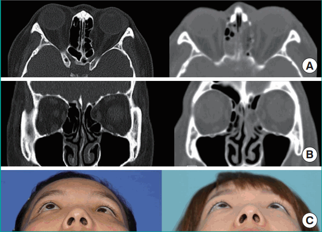
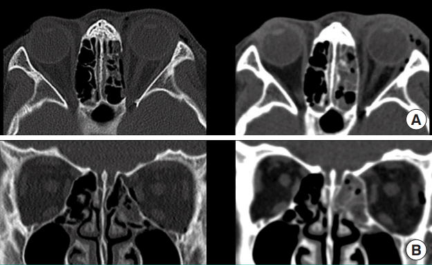
Old fracture case
Computed tomography (CT) scans of the the orbit obtained before and after the operation. The orbital volume of the fractured side was 28.11 cm3 preoperatively and improved postoperatively to 24.89 cm3. The difference on the Hertel scale was 2.02 mm preoperatively, and improved to 0.20 mm postoperatively. (A) Preoperative and postoperative axial CT scans demonstrating that the herniated orbital contents were restored completely. (B) Preoperative and postoperative coronal CT scans demonstrating that the herniated orbital contents were restored completely. (C) Preoperative and postoperative photographs of the patient
Case 2
A 12-year-old male patient visited the emergency room with midfacial trauma resulting from a bicycle accident. The midfacial trauma resulted in a pure medial wall fracture with no other concomitant midfacial fractures. At the time of the visit, he had no functional problems related to diplopia, impaired vision, or abnormal ocular movements. The medial orbital wall fracture and herniated orbital contents on the left side were identified on a CT scan. He underwent an operation using the push-out technique. The herniated orbital contents and the fractured bone segment were restored to the original anatomical position. Porous polyethylene implants were placed in the ethmoidal sinus layer by layer. Postoperative CT imaging revealed an adequately reconstructed medial orbital wall (Fig. 3). No complications were observed.
Acute fracture case
Computed tomography (CT) scans of the orbit obtained before and after the operation. The orbital volume of the fractured side was 28.65 cm3 preoperatively, and improved to 25.10 cm3 postoperatively. The difference on the Hertel scale was 1.65 mm preoperatively, and improved to 0.30 mm postoperatively. (A) Preoperative and postoperative axial CT scans demonstrating that the herniated orbital contents were restored completely. (B) Preoperative and postoperative coronal CT scans demonstrating that the herniated orbital contents were restored completely.
DISCUSSION
The symptoms of medial orbital wall fractures are usually less severe than those of inferior wall fractures because less muscle incarceration takes place and the bony structure is multiply overlapped [5]. Since medial orbital wall fractures are often asymptomatic, they have received less attention in the literature [6,7]. However, they may cause complications such as diplopia, enophthalmos, and the entrapment of extraocular muscles [8,9]. In particular, enophthalmos may not appear immediately after the trauma because soft tissue swelling can last weeks or months. The key to preventing such complications is not only to make an accurate diagnosis, but also to reconstruct the fractured wall of patients in whom surgery is indicated. CT scans enable more accurate diagnoses of orbital fractures, especially the medial wall type [8,10]. Surgical correction is generally necessary in patients with the following symptoms and signs: diplopia lasting over a week, limited EOM, blurred vision resulting from optic nerve compression, a bone deficit greater than 2 cm, or enophthalmos resulting from orbital tissue herniation [11].
The medial orbital wall is formed by the lamina papyracea of the ethmoid, lacrimal, sphenoid, and frontal bones, along with the anterior and posterior ethmoidal arteries, the optic nerve, and other structures. The anatomical complexity of the periorbital area is challenging for surgeons and has spurred a constant search for less invasive methods. To improve surgical outcomes and to reduce complications, various techniques for reconstructing the medial orbital wall have been introduced. The inlay implanting technique, in which multiple porous polyethylene implants are inserted into the ethmoid sinus, has been reported to lead to successful outcomes with few complications. However, when confirming the defect margin of medial orbital wall fractures and detaching the bone and soft tissue around the pathologic area, complications remain possible, as when the conventional method is used. In cases of post-traumatic enophthalmos caused by a medial orbital wall fracture, adhesion of the bone, periosteum, and soft tissue makes the dissection of the fractured bone segment difficult.
The purpose of this study was to develop a minimally invasive and simple surgical method with a reduced range of dissection. Without dissecting and removing the fractured bone segments by excessive force, herniated orbital tissue alongside the fractured bone can be repositioned just by “pushing out.” We were able to reduce the operating time and did not have to worry about complications caused by dissection around the fracture site, such as bleeding, nerve injuries, and soft tissue injuries.
Only one patient underwent a revisional operation immediately after the operation because of newly developed diplopia. He had a comminuted fracture, and a tiny fractured segment was located toward the medial orbital wall, with an orientation that was almost vertical. We missed it on preoperative CT scans. During repositioning of the fractured bony wall, the medial rectus muscle was pricked by the fractured segment. We found the problem on immediate postoperative CT scans, and removed the segment in a revisional operation. The patient who underwent the revisional operation showed improved diplopia symptoms.
In cases of the late correction of old medial orbital wall fractures with post-traumatic enophthalmos, the push-out technique is useful because the mucous membrane surrounding the fractured orbital wall is intact, making the reduction of a single segment more feasible. By using the push-out technique, a surgeon can avoid unnecessary subperiosteal dissection, which may be dangerous due to extensive scar tissue and bleeding from fibrovascular sources. The push-out technique is also indicated for greenstick fractures in young, soft bone in which the ethmoid bone bends and breaks. The bone segments involved with a greenstick fracture are commonly larger and have a low risk of being broken during the push-out technique. The push-out technique is useful for single-hinged trapdoor-type fractures when soft tissue is not entrapped.
Notes
No potential conflict of interest relevant to this article was reported.
Acknowledgements
This study was supported by a 2016 Yeungnam University Research Grant.
Notes
PATIENT CONSENT
The patient provided written informed consent for the publication and the use of their images.