Utility of topical epinephrine for determining the resection range of eyelid sebaceous carcinoma with dermatitis
Article information
Although sebaceous carcinoma is a relatively rare malignant tumor of the skin, death from metastasis can occur in up to 6% of all cases [1]. The first-line treatment is extended surgical resection with appropriate margins. However, if the tumor is associated with dermatitis, its border may be unclear, which can make determination of the resection range difficult. The authors encountered an 86-year-old woman with a right upper eyelid sebaceous carcinoma misdiagnosed as blepharitis, which was treated with a topical steroid ointment for more than 1 year. When the authors operated, rebound inflammation due to the sudden discontinuation of long-term steroid treatment appeared around the tumor, obscuring its border (Fig. 1). However, topical epinephrine (1:100,000 dilution) applied to stop bleeding from the biopsy site incidentally clarified its border, facilitating determination of the appropriate resection range (Fig. 2). Pathological diagnoses of the skin margin using paraffin sections were negative.
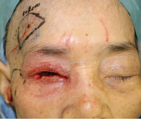
Immediately after lower right eyelid punch biopsy. The border of the tumor was unclear because of the surrounding dermatitis.
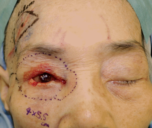
Right eyelid after topical epinephrine (1:100,000 dilution). The border of the tumor was clarified after removal of the epinephrine-soaked gauze. The extent of skin resection was determined (10 mm margin).
Inflammation has five signs: redness, heat sensation, swelling, pain, and dysfunction. Redness and heat sensation due to inflammation arise as a result of capillary dilation and increases in local blood flow. Because epinephrine acts on the α1 receptor and constricts peripheral vessels in the skin, it reduces bleeding from surgical wounds and is therefore useful in surgery [2,3]. It is believed that epinephrine treatment temporarily improved the redness and clarified the tumor border.
In future studies, we would aim to increase the number of cases, including not only eyelid and sebaceous adenocarcinoma, but also other anatomical locations and tumors. The patient consented to publish her clinical course and photographs.
Notes
No potential conflict of interest relevant to this article was reported.
Patient consent
The patient provided written informed consent for the publication and the use of her images.