Treatment of refractory groin lymphocele by surrounding supermicrosurgical lymphaticovenous anastomosis
Article information
Lymphocele is a localized lymph collection that is usually secondary to lymphatic network damage. Large lymphoceles may lead to chronic pain and infection. Herein, we report a case of refractory groin lymphocele treated by supermicrosurgical lymphaticovenous anastomosis.
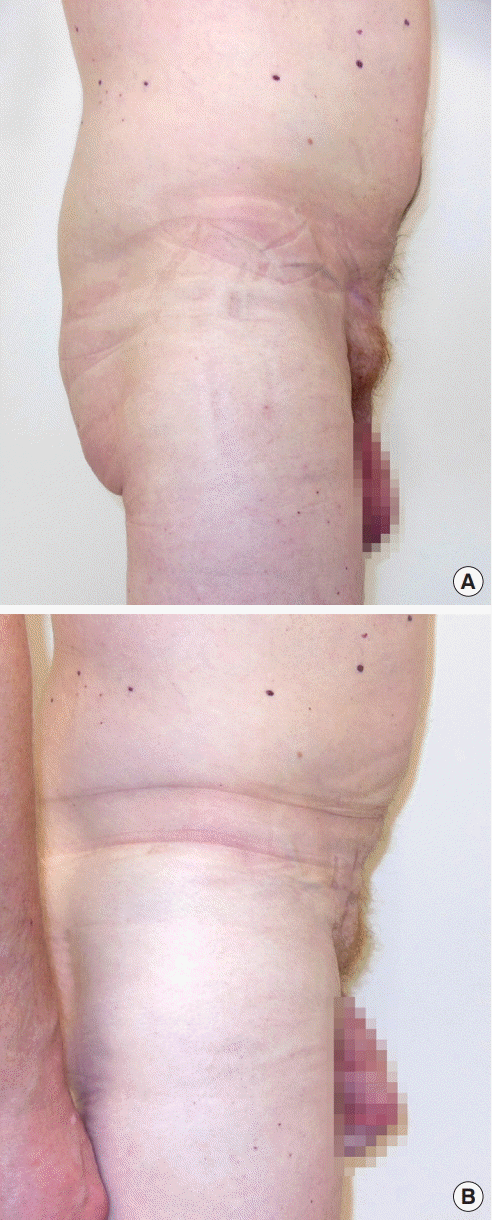
A 43-year-old man had been treated 2 years previously for an inguinal hernia. A few days after surgery, a subcutaneous lymphocele occurred in the groin area (Fig. 1A). The patient presented to our department after the failure of conservative treatment.

Comparison of the patient’s clinical appearance before and after surgery. (A) Preoperative view: a 43-year-old man presented with a refractory groin lymphedema secondary to inguinal hernia surgery. (B) Postoperative view: complete lymphocele volume reduction was visible 3 months after surrounding lymphaticovenous anastomosis.
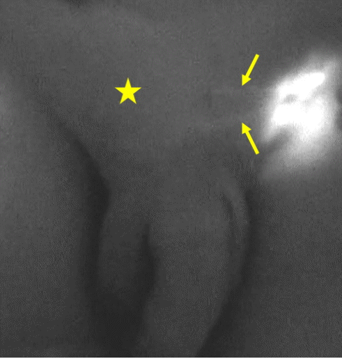
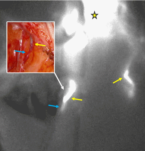
Lymphaticovenous anastomosis under local anesthesia was chosen as a minimally invasive procedure. Magnetic resonance imaging (MRI) was performed preoperatively to delineate the lesion and identify adjacent venules. Indocyanine green fluorescence lymphangiography with a PhotoDynamic Eye (Hamamatsu Photonics Co., Hamamatsu, Japan) was used to visualize the functional lymphatic vessels surrounding the lymphocele area intraoperatively (Fig. 2). Two end-to-end lymphaticovenous anastomoses were performed under high magnification (×27) with 12-0 nylon sutures (Fig. 3). Microvascular anastomosis patency was checked intraoperatively by fluorescence lymphangiography, for which 0.1 mL of indocyanine green was injected inside the lymphocele. A skin massage was performed to diffuse the tracer into the surrounding lymphatic vessels. Positive patency was visualized in each lymphaticovenous anastomosis (Fig. 3).

Intraoperative view of the indocyanine green fluorescence lymphangiography before lymphaticovenous anastomosis. A subdermal injection of 0.1 mL of indocyanine green was made 5 cm medially from the lymphocele (yellow star). Two functional lymphatic vessels (yellow arrows) were visualized through the skin. They ran towards the collection of fluid in the groin (yellow star), which was slightly fluorescent.

Intraoperative view of lymphaticovenous anastomosis patency by indocyanine green fluorescence lymphangiography. An injection of 0.1 mL of indocyanine green was made inside the lymphocele (yellow star). A skin massage was performed to diffuse the tracer into the surrounding lymphatic vessels (yellow arrows). The tracer diffused into the venule (blue arrows).
The patient’s postoperative course was uneventful. The subcutaneous lymphocele volume started to reduce 5 days after surgery. Postoperative MRI confirmed the progressive absorption of the lymph collection. Complete resorption was achieved 3 months postoperatively (Fig. 1B).
Lymphaticovenous anastomoses have been used in lymphocele treatment in a few cases. The technique was successful for pelvic lymphocele [1] and for a localized subcutaneous leg lymphocele after sentinel node biopsy [2]. Lymphatic vessel bypass to a collateral branch of the great saphenous vein was described for postoperative groin lymphocele [3].
To our knowledge, this is the first reported case of refractory groin lymphocele treated by supermicrosurgical lymphaticovenous anastomosis. In contrast to the technique reported by Boccardo et al. [2] and Gentileschi et al. [3], no lymphocele capsule excision was required. The advantage of surrounding lymphaticovenous anastomosis based on small incisions was to minimize the length of surgical scars. Thus, surrounding supermicrosurgical lymphaticovenous anastomosis may be a new option for the management of refractory groin lymphocele.
Notes
No potential conflict of interest relevant to this article was reported.
Patient consent
The patient provided written informed consent for the publication and the use of his images.