An old problem with a new solution: Cost-effective, easy correction of rhinophyma using a disposable razor
Article information
Rhinophyma is the late stage of rosacea, a chronic skin disorder that is characterized by distorting hypertrophy of the sebaceous glands and has been mistakenly connected to alcohol abuse [1], making it a socially stigmatized condition. While patients seek treatment mainly for cosmetic reasons, cases of malignant degeneration have been reported [2]. The treatment of rosacea includes rigorous avoidance of triggers, antibiotics, and retinoids. The mainstay of the treatment of rhinophyma consists of surgery, using a CO2 laser, hydrosurgery, or simple excision. We report the case of a 55-year-old female patient who presented with a case of distorting rhinophyma (Fig. 1) after undergoing conservative treatment with oral isotretinoin and ivermectin without any improvement. Tangential excision was performed using a commercially available, sterile single-blade razor (Fig. 2). Wound dressings using Mepithel (Mölnlycke Health Care, Goeteborg, Sweden) were applied postoperatively. The final aesthetic outcome 10 months postoperatively was pleasing, and the patient’s postoperative course was uneventful. No further corrections were needed. The nose regained a natural contour (Fig. 3). Despite the manifold technologies available for the treatment of rhinophyma, there is no consensus on the best technique. The feasibility of obtaining appropriate histopathological samples is the main advantage of tangential excisional techniques. In such techniques, it is crucial to leave the lower part of the pilosebaceous units intact by careful, step-by-step shaving to guarantee total excision along with undisturbed healing by secondary intention. A sterile, disposable razor is a valuable tool in rhinophyma surgery that is inexpensive, commercially available, and easy to use. This method has been described before [3], but it has largely been forgotten and is not in routine clinical use. Thus, we herein re-describe and recommend this cost-effective technique that unites the advantages of excisional and tangential ablative methods in a single simple-to-perform procedure.
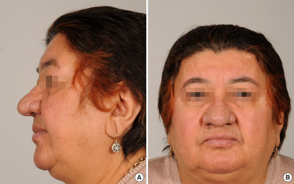
Preoperative lateral (A) and frontal (B) standardized photographs of the patient.
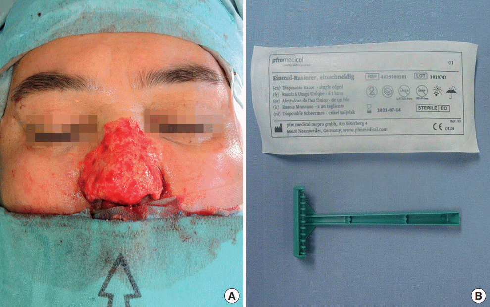
Intraoperative view of the patient (A) operated on with a sterile disposable razor (B).
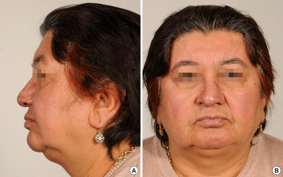
Lateral (A) and frontal (B) photographs of the patient 10 months postoperatively.
Notes
No potential conflict of interest relevant to this article was reported.
Ethical approval
The study was performed in accordance with the principles of the Declaration of Helsinki. Written informed consent was obtained.
Patient consent
The patient provided written informed consent for the publication and the use of her images.