Eruption of a venous malformation through an iliac bone harvesting site after trauma
Article information
Abstract
Harvesting grafts from the anterior iliac bone has been associated with various complications. A 50-year-old woman presented to our department with a chief complaint of right inguinal swelling and pain. Autologous bone grafts had been harvested on two previous occasions from the right anterior iliac crest for use in the reconstruction of multiple facial fractures. Computed tomography and magnetic resonance imaging revealed a full-thickness bone defect in the right anterior iliac crest. A mass was noted in the right gluteus minimus, while a multilocular cystic mass extended from the right iliac crest defect to the right inguinal region. Both the inguinal mass and gluteal mass were removed under general anesthesia. Following histopathological analysis, the gluteal mass was diagnosed as a venous malformation(VM). Based on the patient’s clinical course, iliac bone graft harvesting and trauma to the gluteal region triggered hemorrhaging from the VM. Blood components leaked out from the fragile portion of the iliac bone defect, forming a cystic lesion that developed into the inguinal mass. In this case, a coincidental VM resulted in a rare complication of iliac bone graft harvesting. These sequelae could have been avoided by planning for more appropriate ways to collect the grafts.
INTRODUCTION
Autogenous bone grafts are most commonly harvested from the anterior iliac crest for use in orthopedic surgery, neurosurgery, and oral and maxillofacial surgery. Harvesting grafts from the anterior iliac bone has been associated with various complications, such as persistent pain, hematoma, seroma, nerve injury, wound infection, fractures, pseudoaneurysm, and herniation [1-3]. A range of factors, such as the iliac bone region, bone defect size, patient age, and body type, can affect the likelihood of complications [1,4,5]. However, it is unlikely for a separate lesion to affect the iliac bone harvest site. The authors experienced a rare case in which hemorrhage from a gluteal venous malformation (VM) extended into the inguinal region via the iliac bone harvest site.
CASE
A 50-year-old woman presented to plastic and reconstructive surgery department with a chief complaint of right inguinal swelling and pain. Autologous bone grafts had been harvested on two previous occasions from the right anterior iliac crest for use in the reconstruction of multiple facial fractures incurred during a traffic accident 29 years ago. Two months before presentation, the patient accidentally sat on a piece of metal with her right buttock and developed pain in the right pubic region, and a mass appeared in the inguinal region. The pain continued to worsen while the mass grew, leading her to consult the surgery department at our institution.
A contrast-enhanced computed tomography (CT) scan taken upon presentation at the surgery department suggested active hemorrhage in the right gluteal region, and emergency angiography was performed. Active hemorrhage was confirmed at the anastomosis between the right superior gluteal artery branch and the right medial circumflex femoral artery branch. Successful hemostasis was achieved with two attempts at transcatheter arterial embolization (TAE). Despite this treatment, the inguinal mass continued to grow, and the patient was referred to our department.
A physical examination upon the patient’s initial presentation at our department revealed a large, tense mass (30×12 cm) in the right inguinal region (Fig. 1). The only symptom was mild tenderness. No apparent symptoms were noted in the right gluteal area, and palpation revealed no abnormalities. The vital signs and blood laboratory test data were also normal. A CT scan revealed a full-thickness bone defect in the right anterior iliac crest (Fig. 2A). A mass was noted in the right gluteus minimus (Fig. 2B), while a multilocular cystic mass extended from the right iliac crest defect to the right inguinal region (Fig. 2C). Magnetic resonance imaging (MRI) showed that the gluteal lesions were septated, with low signal intensity on T1-weighted images (Fig. 3A), while T2-weighted images and short TI inversion recovery (STIR) images were of iso-signal to high signal intensity (Fig. 3B and C). The inside of the lesions showed high signal intensity in the T1-weighted and STIR images (Fig. 3A and C), and low signal intensity in the T2-weighted images, suggesting hemorrhagic components (Fig. 3B).
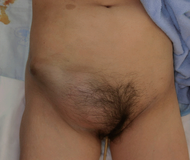
Preoperative photograph
A large, tense mass (30×12 cm) was discovered in the right inguinal region.
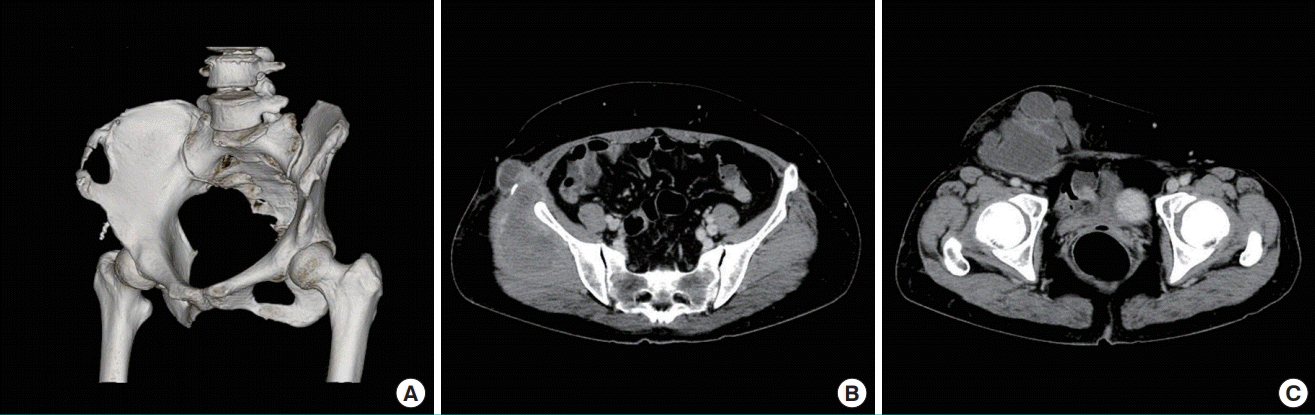
Preoperative computed tomography images
(A) Reconstructed computed tomography image showing a full-thickness bone defect in the right anterior iliac crest. (B) A mass was observed in the gluteus minimus muscle. (C) A multilocular cystic mass covered the area from the right iliac crest defect through the right inguinal region.
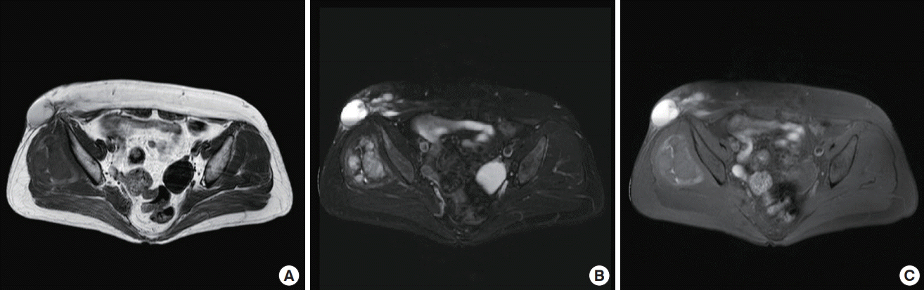
Preoperative magnetic resonance images
The right gluteal mass was composed of septated lesions with low signal intensity on T1-weighted images (A), and high signal intensity on T2-weighted and short TI inversion recovery (STIR) images (B, C). T1-weighted and STIR images suggesting hemorrhagic components inside the lesion showed high signal intensity (A, C), and the corresponding T2-weighted finding was low signal intensity (B).
These imaging findings led us to suspect that the gluteal mass was a VM. The inguinal cystic lesions were suspected to be the result of inflow from the gluteal mass. With localized treatment comprising paracentesis and compression therapy, there was a high risk of recurrence. Since the patient had a history of TAE, we considered surgical resection to be a viable option, and decided to excise the inguinal and gluteal masses simultaneously. Both the inguinal mass and gluteal mass were removed under general anesthesia. A skin incision made immediately below the inguinal mass revealed a multilocular cystic mass on the inguinal ligament that extended from the iliac bone defect to the gluteal region (Fig. 4). This mass entered the gluteal region and formed another mass inside the gluteus minimus muscle. The border separating the gluteal mass from the surrounding tissue was distinct, and it was possible to excise it together with the inguinal lesion. Upon rupture of its membrane, the inguinal lesion produced a dark brown serous exudate (Fig. 5).
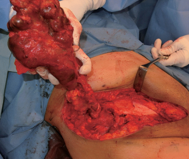
Intraoperative photograph
A multilocular cystic mass was noted on the inguinal ligament. A continuous mass was observed, extending from the iliac bone graft harvesting site to the gluteal site.
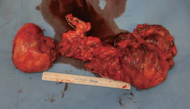
Intraoperative specimen
The inguinal mass was connected to the gluteal mass. Rupturing the membrane on the inguinal mass produced a dark brown exudate.
Histopathological analysis revealed that the inguinal mass had no capsular epithelium, and numerous histiocytes and inflammatory cells covered the surface. Fibrin had adhered to the lumen. The gluteal mass was badly damaged, which we thought to be a result of the presurgical TAE, so it was difficult to determine the overall characteristics of the tumor. Most of it was made up of organized cysts and hematoma, but it also included an area of heterogeneously distributed, irregularly dilated, venous vasculature. Immunostaining of the vascular endothelium was positive for CD34 and negative for D2-40 (Fig. 6). These results were compatible with a VM. Six months after surgery, the patient had recovered with no signs of recurrence (Fig. 7).
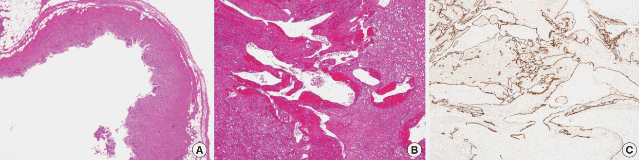
Histopathologic and immunohistochemical findings
(A) The inguinal mass showed no signs of a capsular epithelium, and the surface was covered with multiple histiocytes and inflammatory cells. Fibrin adhered to the lumen (hematoxylin and eosin [H&E], ×20). (B) The gluteal mass revealed heterogeneously distributed, unevenly dilated, venous vasculature (H&E, ×20). (C) Immunostaining of this vascular endothelium was positive for CD34 (×20).
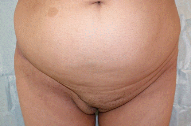
Postoperative photograph
Six months after surgery, the patient had recovered with no signs of recurrence.
DISCUSSION
The complications reported after iliac bone graft harvesting include persistent pain, hematomas, seromas, nerve injuries, infections, and fractures [1]. However, few papers have reported the formation of masses as a complication of iliac bone harvesting. Pseudoaneurysm has, however, been reported after anterior iliac bone graft harvesting [2,3]. In our case, the gluteal mass formation was believed to be a complication of iliac bone harvesting, and pseudoaneurysm was originally suggested in the differential diagnosis. However, the cases of pseudoaneurysm reported after anterior iliac bone graft harvesting were of deep circumflex iliac artery origin [2,3], which was inconsistent with the site of hemorrhage in the present case (Fig. 1). Ultimately, the MRI results led us to reject the diagnosis of pseudoaneurysm and to suspect a VM; the final diagnosis of VM was based on histopathology.
VM is the most common vascular developmental anomaly (birth defect). It produces slow-flow pooled blood lesions with vein-like lumen proliferation. These defects are caused by developmental arrest of the venous system during various stages of embryogenesis [6]. They do not disappear spontaneously, and symptoms can progress or become aggravated with growth or surgical stimulation.
Based on the above considerations, the gluteal VM must have been present even before iliac bone graft harvesting. It was a coincidence that a VM had formed in the gluteal area on the same side as the graft harvesting. We speculate that the VM was asymptomatic because it was in the gluteus minimus muscle. The problem was that the gluteal VM extended into the inguinal region. In this case, a full-thickness bone defect was present because the iliac bone had been harvested multiple times. Clearly, this is one of the fundamental reasons why the tumor extended into the inguinal region. Based on this patient’s clinical course, we speculate that an arteriovenous shunt formed in the VM because of active hemorrhage in the right gluteal region, as shown by emergency angiography, and that the arteriovenous shunt was caused by multiple rounds of iliac bone graft harvesting.
Furthermore, trauma to the gluteal region triggered an increase in the arteriovenous shunt blood flow and an increase in the tumor’s internal pressure. Finally, blood components leaked out from the fragile portion of the iliac bone defect, forming a cystic lesion that developed into the inguinal mass. We speculate that this is another reason why the tumor extended into the inguinal region.
To summarize, the pathogenesis in this case was a coincidentally present VM, multiple surgical procedures in the same region, and full-thickness iliac bone graft harvesting.
Analogously, incisional hernias are complications that occur after surgical invasion and full-thickness iliac bone graft harvesting. Hernias after iliac graft collection were first reported by Oldfield in 1945 [7]. This complication occurs in 5% of cases after bone harvesting from the iliac crest, according to Auleda et al. [8]. Risk factors include large bone defects, full-thickness graft harvesting, advanced age, obesity, and poorly-developed musculature [5,9]. In addition, Hamad and Majeed [5] recommended avoiding the harvest of full-thickness grafts when partial-thickness grafts suffice.
In this case, the tumor extending into the inguinal region could have been prevented by leaving either the inner or outer plate intact during iliac bone graft harvesting instead of collecting fullthickness slices. However, Ahlmann et al. [4] investigated harvest-site morbidity in 88 patients and concluded that while anterior iliac crest harvesting was associated with an 8% complication rate, complications were seen in only 2% of cases where the posterior iliac crest was harvested. In cases such as this patient, where multiple bone grafts are required, the surgeon should consider harvesting from the posterior iliac crest.
In this case, a coincidental VM resulted in a rare complication of iliac bone graft harvesting. However, these sequelae could have been avoided by planning for more appropriate ways to collect the grafts.
Notes
No potential conflict of interest relevant to this article was reported.
Ethical approval
The study was performed in accordance with the principles of the Declaration of Helsinki. Written informed consent was obtained.
Patient consent
The patient provided written informed consent for the publication and the use of her images.