Vastus intermedius–anterolateral thigh chimeric free flap for lower extremity reconstruction
Article information

Generally, for the chimeric anterolateral thigh (ALT) flap, the vastus lateralis (VL) muscle is simultaneously harvested with a skin paddle [1,2]. However, it may be difficult to use an ALT chimeric flap including the VL if the distance between the defects is too great.
The vessels that flow into the VL are located near the perforator that runs from the descending branch of the lateral circumflex femoral artery (LCFA-db) to the skin paddle. On the other hand, the vastus intermedius (VI) is a type II muscle, for which the dominant pedicle is supplied by the LCFA-db and the minor pedicle is supplied by the profunda femoris and superficial femoral arteries. Moreover, the main pedicle branches from a site approximately 7-cm distal to the site where the descending branch originates from the LCFA and enters the VI, while small vessels from a more distal descending branch supply blood to the muscle [3]; thus, a chimeric flap that includes the VI muscle can be an option in the reconstruction of two defects far from each other.
A 65-year-old man had two defects (17×8.5 cm2, 7×3.5 cm2) on the distal tibia and lateral foot. The distance between the two defects was 9 cm (Fig. 1). We planned a chimeric free flap intraoperatively. After elevation of a fasciocutaneous flap, a branching point at approximately 3.5 cm distal to where the LCFA-db originated from the VI was identified, after which a muscle flap was elevated (Fig. 2). The pedicle was microanastomosed to the anterior tibial vessels. The muscle component was covered by skin grafting and the donor site was primarily closed. The patient was followed up for 6 months after surgery, during which time he showed good recovery with no wound problems or contour abnormalities (Fig. 1B).
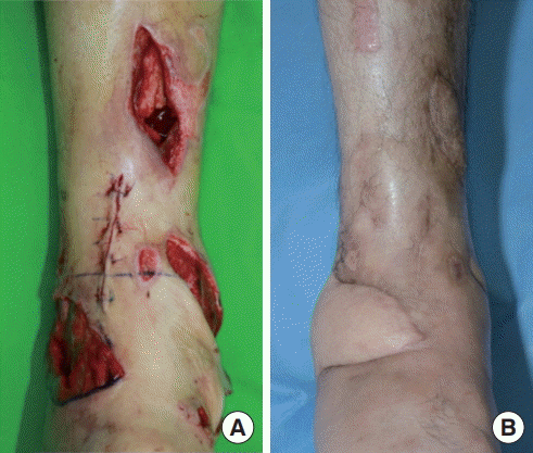
There were two defects on distal tibia and lateral foot, approximately 9 cm apart. (A) An open wound (17×8.5cm2) including necrotic tissue on the lateral side of foot, another open wound with bone exposure (7×3.5 cm2) on the superomedial side of right distal pretibia. (B) Six months postoperatively after the vastus intermediusanterolateral thigh chimeric free flap.
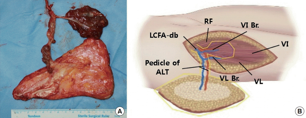
(A) Elevated vastus intermedius-anterolateral thigh (VI-ALT) chimeric free flap. (B) Scheme of the VI-ALT chimeric free flap. RF, rectus femoris; VL, vastus lateralis; VL Br., branch of the VL; VI Br., branch of the VI; LCFA-db, descending branch of the lateral circumflex femoral artery.
The VI muscle–ALT chimeric free flap was able to achieve favorable outcomes with minimal donor site morbidity in the reconstruction of multiple defects that are far apart.
Notes
No potential conflict of interest relevant to this article was reported.
Ethical approval
The study was performed in accordance with the principles of the Declaration of Helsinki. Written informed consent was obtained.
Patient consent
The patient provided written informed consent for the publication and the use of his images.