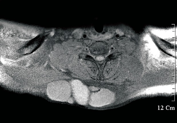
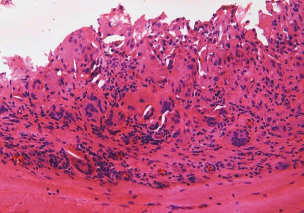
We appreciate Dr. Yu Jin Kim for her insightful commentary on our recent article [1]. The additional masses that are mentioned in Dr. Yujin Kim's commentary were attached to the two masses shown on the left side of the gross specimen figure, each with its own epidermal layer [1,2]. The epidermal cyst in the center of the coronal computed tomography image is that which had the foreign body reaction noted by histology (Figs. 1, 2). Likewise, this same cyst is the one that was found to be mildly warm during pre-operative clinical examination.
Our statement that cites Reference 7 was intended to describe skin appendage tumors as having pathologic findings of nevus comedonicus [1-3]. We agree with the opinion that your article should have been more appropriately referenced in the discussion about direct pathologic findings and surgical treatments. Your advice is acknowledged and appreciated.
In the present case report, we tried to present the possibility that nevus comedonicus in the shallow epidermal layer can progress to a subcutaneous giant epidermal cyst as a late complication. In other words, nevus comedonicus should be considered in the differential diagnosis of other deep subcutaneous tumors.
Conservative treatments are aimed at reducing the formation of comedones and/or controlling infection. Considering surgical treatment, ordinary excision can be performed on localized lesions, including small pits [3,4]. In the case of an extensive lesion, wide excision and tissue expander reconstruction may be used [5]. In the present case, the dilated comedone was large enough to be explored by a probe. During exploration, the comedones seemed to have pits up to 1 cm in size, thus a wide excision with a 1 cm margin was selected in our case. Finally, we believe that the treatment modality chosen should be based on the level of the patient's discomfort, in addition to aesthetic concerns, scar formation, abscesses, and epidermal inclusion cysts.