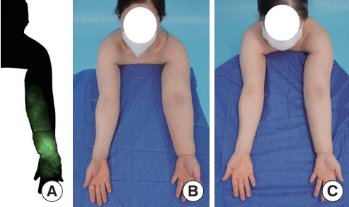
Vascularized lymph node transfer (VLNT) and lymphovenous anastomosis (LVA) can be performed simultaneously or independently, depending on the patient’s lymphedema stage [1]. A 54-year-old woman underwent bilateral total mastectomy in 2016, with sentinel lymph node biopsy for the right breast and axillary lymph node dissection for the left breast, followed by chemoradiotherapy. The patient developed progressive lymphedema (indocyanine green dermal stage IV–V), and LVA and VLNT were simultaneously performed (Fig. 1).
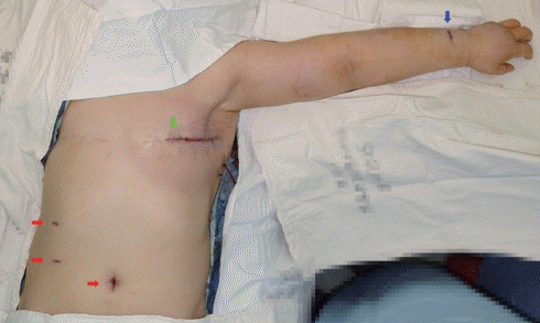
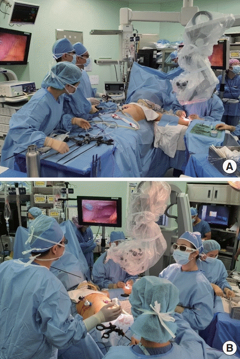
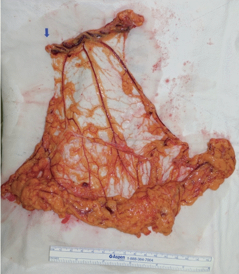
The patient was laid supine and draped as illustrated in Fig. 2. First, the recipient vessels (thoracodorsal artery and vein) were prepared and complete scar tissue excision of the axilla and lateral chest was performed. A general surgeon then laparoscopically harvested an omental flap while a plastic surgeon performed LVA. With well-positioned monitors and microscope (Fig. 3), both the harvest and LVA were performed without interfering with each other’s operative field (Fig. 4). The upper part of the flap, with gastroepiploic lymph nodes, was inset in the axilla, and the remnant omental tissue was inset in the lateral chest.
The advantages of the omental flap are minimal donor-site morbidity (e.g., iatrogenic lymphedema), the large diameter of the gastroepiploic vessels, and the potential for omental tissue to absorb lymphatic fluid [2]. Additionally, with the two-team approach, the overall operative time can be reduced by 1–3 hours compared to when other donor sites, such as the groin or submental space, are used. The patient reported improvement in swelling 2–3 days postoperatively and demonstrated fewer episodes of cellulitis and pruritis, along with a reduction in limb volume by approximately 20%, at 85 days postoperatively (Fig. 1).