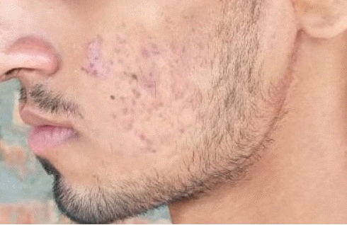
Venous malformations (VMs) of the head and neck region are a common condition that occurs due to abnormal vascular morphogenesis. Typically, VMs are soft and compressible. The common symptoms include low self-esteem due to an unsightly appearance, pain because of thrombosis, and rarely, palpable phleboliths. Phleboliths result from the stagnation of blood in these low-flow malformations, causing a thrombus and progressive lamellar fibrosis, followed by mineralization. We present a case of a VM on the cheek with multiple large palpable phleboliths.
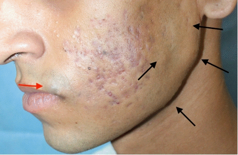
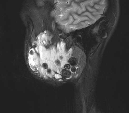
Our 21-year-old patient presented with facial asymmetry and irregularity on the left side of the face since childhood (Fig. 1). He was diagnosed with a large VM of the left side cheek in the masticator space, buccal space, infra-temporal fossa, and left parotid and submandibular spaces with multiple phleboliths (Fig. 2).
The optimal treatment for VM would be complete excision of the lesion to avoid any recurrence [1]. However, in this patient, excision could have resulted in a deformity worse than the malformation. The accepted first-line treatment of problematic VMs is sclerotherapy [2], but a palpable phlebolith remains unaffected by sclerotherapy and could be an indicator for surgery.
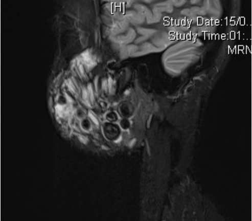
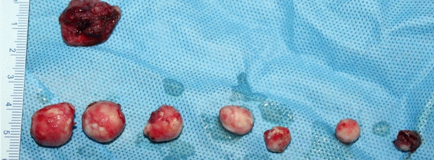
Considering the diffuse nature of the malformation in the cheek, with a high risk of iatrogenic facial nerve injury and bleeding with surgery, this patient was advised to undergo sclerotherapy initially. The commonly used agents in sclerotherapy include sodium tetradecyl sulfate (STS) and ethanol [2]. Since STS is a safer agent in the vicinity of the nerve, three sclerotherapy sessions with 16, 14, and 9 mL of intralesional STS each were done in the interventional radiology suite. One year after the last session of sclerotherapy, a repeat magnetic resonance imaging scan demonstrated a marginal decrease in size of the lesion in addition to decreased intensity when compared to imaging done 2 years previously, suggestive of scar tissue following sclerotherapy. However, predictably, there was no change in the size of phleboliths (Fig. 3). Surgery was planned for excision of phleboliths, not the VM, given its diffuse nature and potential risk of injury to the facial nerve. We successfully removed seven phleboliths of different sizes through a pre-auricular incision, the largest being 1.5 × 1.5 cm, without significant blood loss or any facial nerve injury (Fig. 4). Another solitary VM on the upper lip was excised, and primary closure was done. Postoperatively, the patient was advised to wear a chin strap for compression. At a 4-month follow-up visit, the patient was satisfied with a smooth surface on the cheek (Fig. 5).
Multiple palpable phleboliths in a VM of the head and neck are an uncommon indication for combination treatment using sclerotherapy and surgery, which has not yet been discussed in the literature. There have been reports of patients with individual palpable phleboliths of the maxillofacial region, which have been excised, or multiple small phleboliths for which only sclerotherapy was indicated [3]. This is an unusual case presentation, wherein a surgical intervention was indicated for the deformity due to phleboliths. Using sclerotherapy before surgery was beneficial by minimizing intraoperative bleeding, thereby enabling a reasonably clean dissection that avoided any inadvertent nerve injury and led to a satisfactory outcome.


