Occipital artery pseudoaneurysms are rare vascular lesions, and are most often reported as a consequence of trauma [12]. We report the rare case of a spontaneous pseudoaneurysm of the proximal occipital artery that presented as a neck mass.
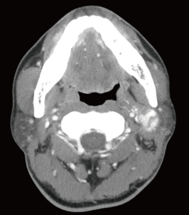
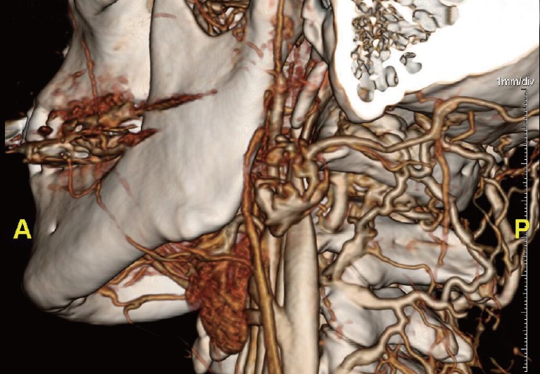
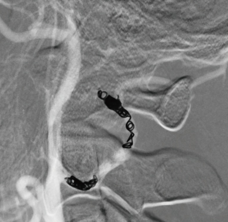
A 44-year-old male patient presented with a pulsatile swelling lesion in the neck that had grown spontaneously over the course of a year (Fig. 1). The swelling had gradually progressed, and he had no previous history of head trauma, infection, or a genetic disorder. The patient was found to have a firm, non-tender, pulsating mass in the left side of the neck that measured 4×3 cm. Ultrasonography showed turbulent flow and dilatation of the involved vessel. Neck computed tomography showed partial opacification of the swelling and revealed a pseudoaneurysm arising from the left proximal occipital artery (Fig. 2). The decision was made to embolize the lesion via angiography (Fig. 3). The mass was treated successfully by endovascular coil embolization (Fig. 4). One year postoperatively, the swelling had improved and recurrence was not detected.
Most occipital artery pseudoaneurysms are consequences of trauma, or are secondary to infections or a genetic disorder [3]. There have been no previous reports of a spontaneous pseudoaneurysm of the occipital artery presenting as a neck mass. The diagnosis can be made based on a thorough history, the exclusion of other possibilities, and imaging studies. Currently, computed tomography angiography is regarded as the most definitive noninvasive diagnostic tool. The advantages of treatment include a reduced risk of hemorrhage, pain relief, and alleviation of cosmetic disfigurement. Treatment options include surgical excision, ligation of the feeding artery, trapping of the pseudoaneurysm, and endovascular arterial embolization and coil occlusion [3].