Lee and Kim: Reconstruction of the Alar-Facial Groove Using a Nasolabial Flap and Medial Directional Force with a ‘Tissue-Adding’ Effect
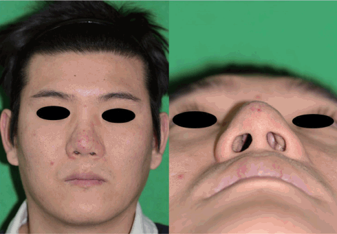
Reconstructing the nose, especially the alar-facial groove, is difficult because of its 3-dimensional structural characteristics. We report the case of a 33-year-old man with a history of crush injury to the nose 15 years previously. We performed reconstruction because of scar contracture formation in the left alar-facial groove (Fig. 1).
This study was reviewed and approved by the Ethics Review Board of the Inje University Health Center.
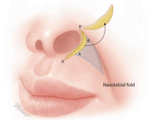
A V-Y advancement flap was designed by setting the nasolabial fold as the superior margin and the elevated alar-facial groove as the medial margin. A cutaneous perforator flap was then elevated [1]. The scar tissue in the alar-facial groove, including the skin and subcutaneous layer, was minimally excised, by 1.0×0.2 cm (Fig. 2).
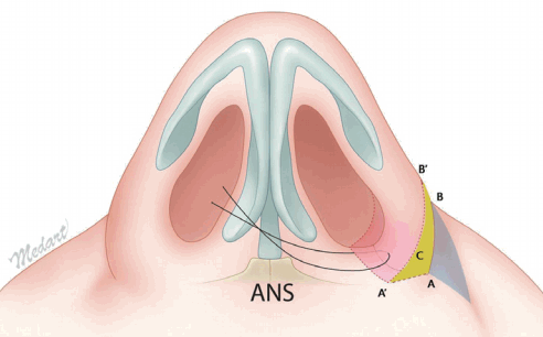
The septum was peeled back to expose the anterior nasal spine, and the bottom surface of the alar side was fixed to a firm area near the anterior nasal spine. This can be done via open rhinoplasty or a minimal incision in the mucosa inside the nostril (Fig. 3).
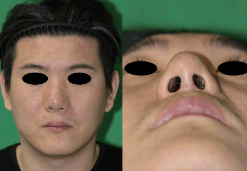
The alar-side surface of the area from which the scar tissue was excised and the medial area of the nasolabial V-Y flap were sutured together. In this manner, a stronger and more prominent secondary alar-facial groove was constructed (Fig. 4).
The definitive treatment for patients needing alar-facial groove reconstruction has not been established. The skirt flap is not optimal for a prominent alar-facial groove [2], nor is the feather-edge rolled-in flap optimal for resolving the tension around the groove [3]. We used a nasolabial flap and ‘tissue-adding’ to reconstruct the alar-facial groove. This technique reduces tension and yields more prominent results by providing a force in the medial direction.

Notes

No potential conflict of interest relevant to this article was reported.

Notes

PATIENT CONSENT

The patient provided written informed consent for the publication and the use of their images.

Fig. 1.
Preoperative view showing the vague alar-facial groove resulting from a crush injury.
aps-2017-44-5-469f1.tif
Fig. 2.
Illustration of the surgical technique. Scar tissue on the alar-facial groove was resected with a minimal incision and elevated in the nasolabial fold direction with a V-Y flap design. Point A moved to A’, and point B moved to B’ by the V-Y advancement flap.
aps-2017-44-5-469f2.tif
Fig. 3.
Fixation of the alar base, close to the hard area of the anterior nasal spine, where it forms a reentrant alar-facial groove. The location of fixation should be decided based on the symmetry of both sides of the nasal cavity. If only reconstruction of the alar-facial groove is planned, a minimal incision can be made in the mucosa inside the nostril. The yellow (C) area corresponds to excised scar tissue. ANS, anterior nasal spine.
aps-2017-44-5-469f3.tif
Fig. 4.
Postoperative view flap 5 months after surgery showing the formation of the reentrant area on the initially vague alar-facial groove and minimal scarring caused by the V-Y advancement.
aps-2017-44-5-469f4.tif

References

1. Han D, Mangoba DS, Lee D, et al. Reconstruction of nasal alar defects in asian patients. Arch Facial Plast Surg 2012;14:312–7.
crossref pmid
2. Ueda K, Shigemura Y, Hara M, et al. Skirt flap for nasal alar reconstruction. Plast Reconstr Surg Glob Open 2014;2:e157.
crossref pmid pmc
3. Park JL, Oh CH, Hwang K, et al. Correction of an alar web with a feather-edge rolled-in flap. J Craniofac Surg 2014;25:2192–5.
crossref pmid