Park, Kim, Kim, Choi, Yang, and Lee: Irritated Subtype of Seborrheic Keratosis in the External Auditory Canal
Seborrheic keratosis (SK) is commonly observed throughout the body, except the palms and soles [1]. However, SK in the external auditory canal (EAC) is rare [2,3]. In this report, we describe a case of SK in the EAC.
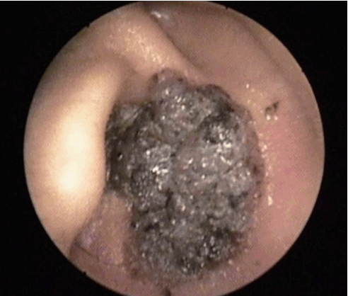
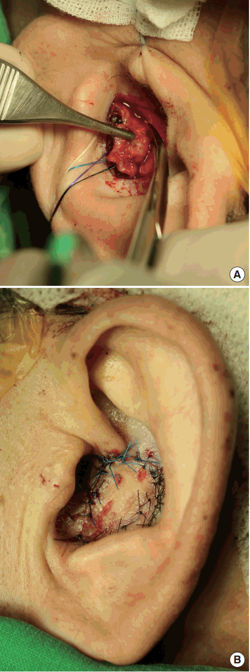
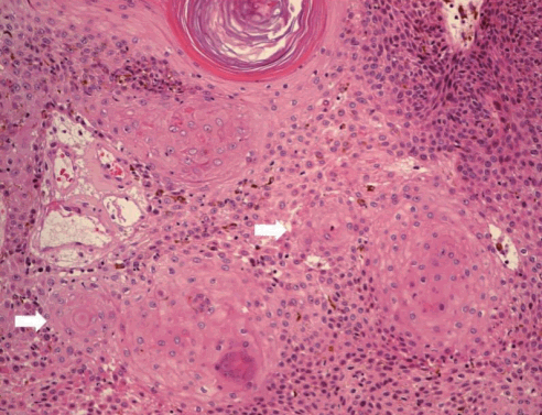
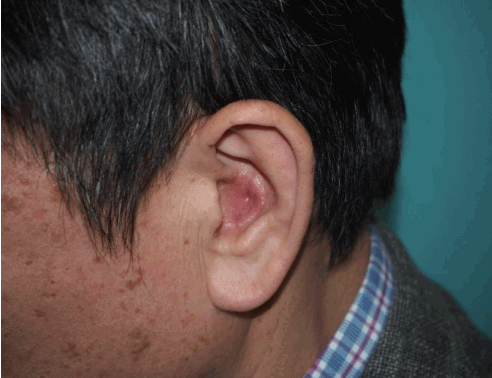
A 56-year-old man presented to our outpatient plastic surgery clinic with a 1-year history of a slow-growing, painless mass in his left auricle. In the physical examination, we observed a 2.5-×2.0-cm blackish papillomatous lesion within the left cavum concha, extending into the EAC (Fig. 1). There was no palpable enlargement of the regional lymph nodes. An incisional biopsy was performed to rule out a malignant skin tumor, and the histopathological examination revealed SK. Subsequently, an excisional biopsy was performed (Fig. 2A). The EAC and cavum concha were reconstructed with a full-thickness skin graft taken from the retroauricular region (Fig. 2B). The second histopathological examination confirmed the final diagnosis of the irritated subtype of SK, without evidence of malignancy (Fig. 3). At a 6-month follow-up visit, no recurrence was noted (Fig. 4).
Histopathologically, SKs are classified into 7 histological subtypes: acanthotic, hyperkeratotic, adenoid, clonal, bowenoid, irritated, and melanoacanthoma [1]. The acanthotic subtype is the most common [1]. However, in our patient, the histopathological examination confirmed the irritated subtype of SK, which rarely arises in the EAC [2]. To our knowledge, only 4 cases of the irritated subtype of SK in the ear have been presented in the English-language literature.
The clinical differential diagnosis of SK is very important because the disorder can be confused with a malignant skin tumor [1,2]. Therefore, a biopsy should always be performed to exclude malignancy.

Notes

No potential conflict of interest relevant to this article was reported.

Notes

PATIENT CONSENT

The patient provided written informed consent for the publication and the use of their images.

Fig. 1.
Otoscopy reveals a blackish papillomatous lesion within the left cavum concha, extending into the external auditory meatus.
aps-2017-01081f1.tif
Fig. 2.
(A) Intraoperative photograph; the seborrheic keratosis within the left cavum concha, extending into the external auditory meatus, was completely excised. (B) Postoperative photograph after excision, showing the defect site reconstructed with a full-thickness skin graft.
aps-2017-01081f2.tif
Fig. 3.
The characteristics of the irritated subtype are as follows: (1) onion-skin-like aggregations of eosinophilic squamous epithelial cells (squamous eddies); (2) inflammatory cell infiltrates with a partly lichenoid appearance in the dermis; and (3) possible appearance of acantholysis, dyskeratosis, spongiosis, and apoptotic basal cells. The photograph shows squamous metaplasia with abundant eosinophilic cytoplasm and whorled squamous eddies (white arrows) in the resected tissue (H&E, ×200).
aps-2017-01081f3.tif
Fig. 4.
Six-month postoperative photograph showing good cosmetic results without recurrence.
aps-2017-01081f4.tif

References

1. Hafner C, Vogt T. Seborrheic keratosis. J Dtsch Dermatol Ges 2008;6:664–77.
crossref pmid
2. Konishi E, Nakashima Y, Manabe T, et al. Irritated seborrheic keratosis of the external ear canal. Pathol Int 2003;53:622–6.
crossref pmid
3. Izquierdo Velasquez JC, Campos Mahecha AM, Duarte Silva JP. Seborrheic keratosis of the external auditory canal. Otol Neurotol 2012;33:e61–2.
crossref pmid