We read with interest the recent review of reconstructive options in upper limb circumferential defects “Soft tissue reconstruction of complete circumferential defects of the upper extremity”, by Ng et al. [1] published in May 2017. It is clear from the dearth of reported cases in the literature that this is a relatively rare, and severe, defect pattern with no clear consensus on the best method of reconstruction. The authors describe a number of grading systems of these injuries, including that by Tajima [2], which describes the depth of injury in relation to structures affected. The authors suggest a treatment algorithm for reconstruction of such defects based on aetiology. The authors correctly point out that in addition to size, choice of reconstruction must take into account volume, mobility, and pliability to maximise functional outcome [3].
Defect aetiology should indeed influence choice of reconstructive option, but operative techniques can be tailored to fill a defect exactly, taking into consideration which structures are exposed: nerve, bone, joint and tendon must be covered with a durable vascularised flap, whereas muscle can be skin grafted. Where possible, flaps overlying tendons should be fascial to maximize glide, as described, for example, by Muneuchi et al. [4] for a defect on the dorsum of the hand. Free flap design can be modified to increase pliability and decrease volume even in areas where circumferential vascularised cover is vital. A case series by Kim et al. [5] describes a ‘super-thin’ flap raised without the adipose layer during flap harvest to increase pliability, while another reports use of a fascial flap in upper limb and digit reconstruction. A combination of such techniques may be necessary in large and complex defects.
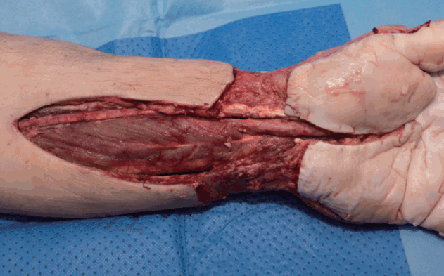
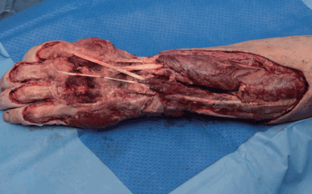
We recently managed a patient following extensive debridement for necrotizing fasciitis of the forearm, wrist and hand. The patient was left with a circumferential skin and soft tissue defect extending from mid-forearm proximally to the dorsal proximal interphalangeal joints (PIPJs) and the volar wrist crease distally. The total size of the defect was 19 cm in circumference by 34 cm in length. Most of the volar forearm defect had healthy muscle at its base, but 8 cm of the median nerve and flexor carpi radialis tendon was exposed at the wrist. Dorsally, all carpal bones and extensor tendons in zones III–VII were exposed (Figs. 1, 2).
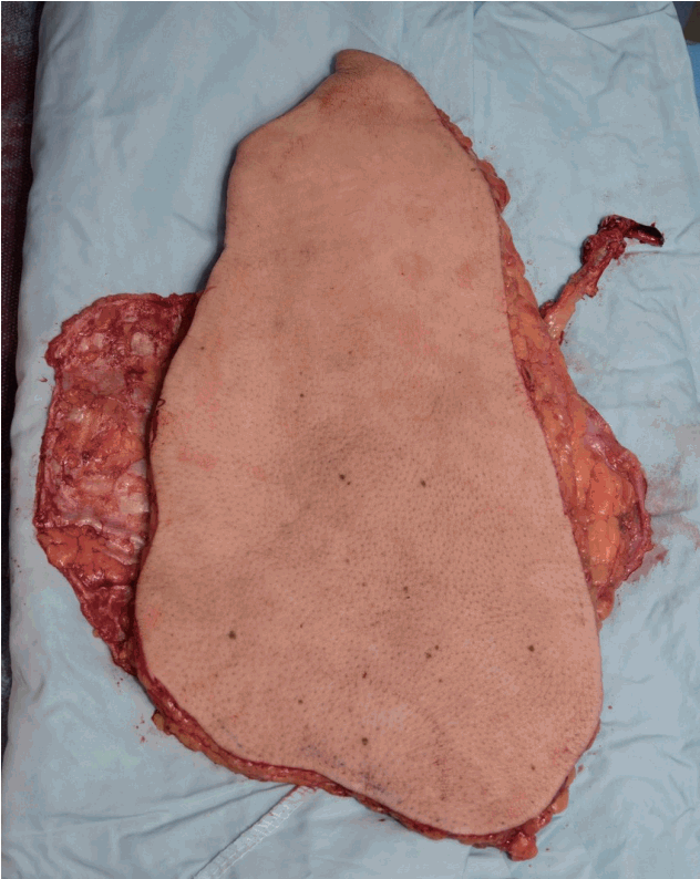
This defect clearly required circumferential durable vascularized tissue cover at the wrist to cover the median nerve volarly and tendons and joints dorsally. Local and regional tissue transfer was not an option due to the extensive nature of the defect. An anterolateral thigh flap was planned, but no suitable perforators were found pre- or intraoperatively so an anteromedial thigh flap was performed. The flap was designed and raised to exactly fit the dorsal defect using a foam template. The fingers were temporarily syndactylised to allow the flap to cover the dorsum of all four fingers to the PIPJs. Fascial extensions were included in the flap design laterally and medially (Fig. 3) and were wrapped around to cover the exposed median nerve at the wrist and split skin grafted. The remaining volar defect was split skin grafted and negative pressure wound therapy was applied for five days to bolster the graft.
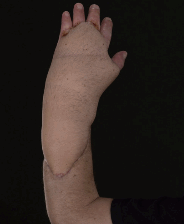
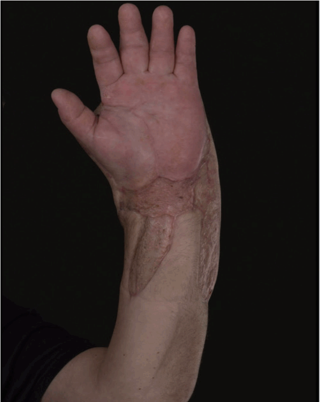
Superficial flap necrosis requiring minimal debridement occurred in the dorsal, distal 5 mm of the flap. The distal 2 cm of one fascial extension required excision and repeat split skin grafting. The rest of the flap survived, as did the split skin grafts (Figs. 4, 5). At four months postoperative, the patient is undergoing intensive physiotherapy. His hand is currently only used as a post, but his range of movement and function is improving each week. He is planned for further surgery to de-syndactylise his fingers, thin the flap, and stabilize the wrist (not done at the time of reconstruction due to risk of infection).
We agree with the authors that these defects are difficult to manage, and achieving limb salvage with acceptable cosmesis and function is a significant challenge. We believe that the aetiology of the injury must be considered, but ultimately the size of defect and structures at its base will determine whether flaps, grafts or a combination is required. The inclusion of fascia-only extensions to cover the volar wrist defect reduced the overall volume of our flap and increased pliability at the wrist joint. This is a useful modification to minimise the bulk of “wrap-around” or “sandwich” flaps, and potentially allow salvage of limbs that would otherwise be unreconstructable.