Extraskeletal soft tissue chondroma (ESTC) is an extremely rare benign hyaline cartilaginous tumor that almost exclusively occurs in the soft tissue of the hands and feet. To correctly diagnose cases of chondroma, clinical, radiological, and cytological investigations are essential [1,2]. An X-ray should be performed as first imaging step, in order to plan surgery precisely. To eliminate bone involvement, magnetic resonance imaging (MRI) is obligatory, and to confirm the diagnosis of ESTC, histological staining should be performed [3].
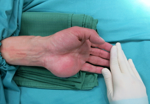
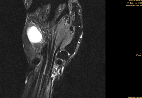
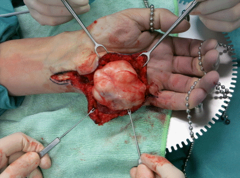
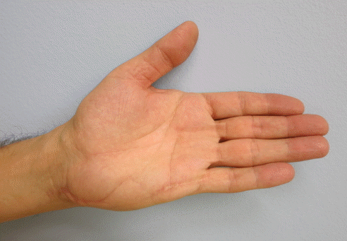
With the patient’s approval, we report the case of a 54-year old man with symptoms of ulnar nerve compression due to a giant ESTC of the left hand (Fig. 1). A physical examination, plain radiographs, and MRI revealed a 4×7 cm soft tissue tumor with hyperintense T2 (Fig. 2) and moderately hypointense T1 MRI signaling located at the hypothenar area with no bone involvement. After verification by histology, excisional biopsy and ulnar nerve decompression were performed (Fig. 3). Pathological examination confirmed the suspected diagnosis of ESTC. The patient’s postoperative recovery was unremarkable and no sign of neurological deficit, pain, or recurrence was observed at follow-up (Fig. 4).
ESTC is a particularly infrequent soft-tissue tumor of the hand. After the diagnosis of ESTC, the first-line therapy is excisional biopsy [4]. The reported local recurrence rates of chondromas are relatively high, so total resection including the capsule should be performed for prophylactic reasons [5]. Since extraskeletal chondromas are often located in close proximity to vulnerable anatomical structures in the hand, it is important to balance radical excision with the preservation of functional structures in this region. Profound hand surgical expertise is needed for the removal of large ESTCs to allow for full functional recovery after tumor resection.